
Sci Rep. 2022;12(1):19638.
Robust R-peak detection in an electrocardiogram with stationary wavelet transformation and separable convolution.
제1저자 윤동환 · 교신저자 한승석
소속 (6)
- Department of Biomedical Sciences, Seoul National University College of Medicine, Seoul, Korea.
- Department of Internal Medicine, Seoul National University College of Medicine, Seoul, Korea.
- Department of Anesthesiology and Pain Medicine, Seoul National University College of Medicine and Seoul National University Hospital, Seoul, Korea.
- Division of Cardiology, Department of Internal Medicine, Seoul National University Hospital, Seoul, Korea.
- Transdisciplinary Department of Medicine and Advanced Technology, Seoul National University Hospital, Seoul, Korea.
- Department of Internal Medicine, Seoul National University College of Medicine, Seoul, Korea. hansway7@snu.ac.kr.
Abstract
R-peak detection is an essential step in analyzing electrocardiograms (ECGs). Previous deep learning models reported their performance primarily in a single database, and some models did not perform at the highest levels when applied to a database different from the testing database. To achieve high performances in cross-database validations, we developed a novel deep learning model for R-peak detection using stationary wavelet transform (SWT) and separable convolution. Three databases (i.e., the MIT-BIH Arrhythmia [MIT-BIH], the Institute of Cardiological Technics [INCART], and the QT) were used in both the training and testing models, and the MIT-BIH ST Change (MIT-BIH-ST), European ST-T, TELE and MIT-BIH Noise Stress Test (MIT-BIH-NST) databases were further used for testing. The detail coefficient of level 4 decomposition by SWT and the first derivative from filtered ECGs were used for model inputs, and the interval of 150 ms centered at marked peaks was used for labels. Separable convolution with atrous spatial pyramidal pooling was selected as the model's architecture, and noise-augmented waveforms of 5.69 s duration (2048 size in 360 Hz) were used in training. The model performance was evaluated using cross-database validation. The F1 scores of the peak detection model were 0.9994, 0.9985, and 0.9999 in the MIT-BIH, INCART, and QT databases, respectively. When the above three databases were pooled, the F1 scores were 0.9993 for fivefold cross-validation and 0.9991 for cross-database validation. The model performance remained high for MIT-BIH-ST, European ST-T, and TELE, with F1 scores of 0.9995, 0.9988, and 0.9790, respectively. The model performance when trained by severe noise augmentation increased for the MIT-BIH-NST database (F1 scores from 0.9504 to 0.9759) and decreased for the MIT-BIH database (F1 scores from 0.9994 to 0.9991). The present SWT and separable convolution-based model for R-peak detection yields a high performance even for cross-database validations.
© 2022. The Author(s).
Figures
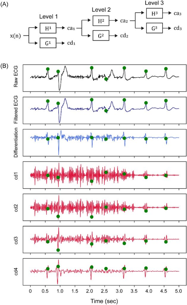
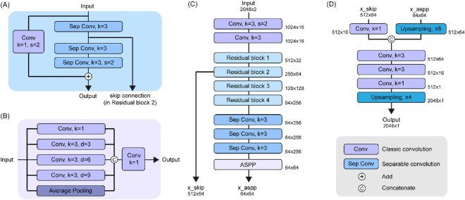
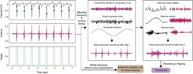
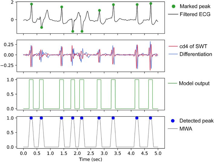
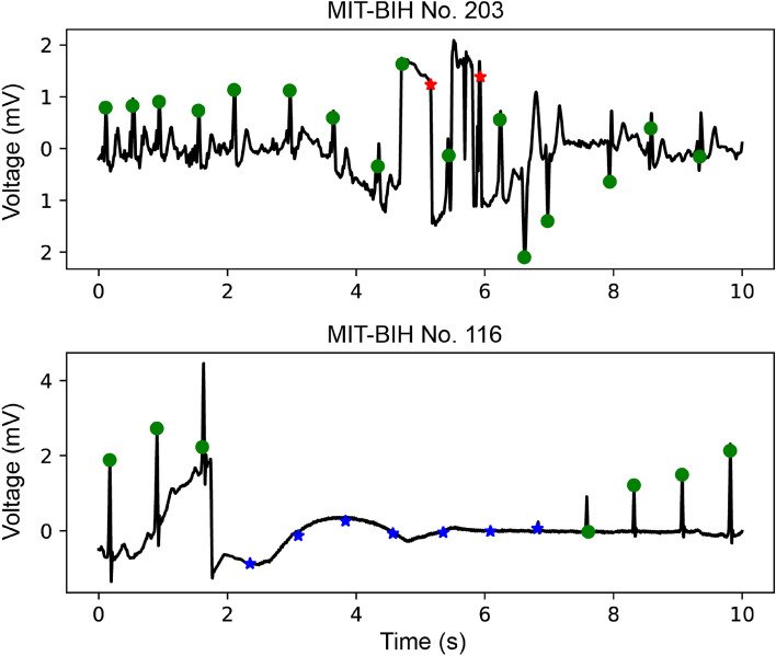
그림 출처: PubMed Central 공개 논문 (PMC9669048)
Conflict of interest statement
The authors declare no competing interests.
그 밖의 정보
- 출판 형태
- Journal Article
- 저널
- Scientific reports · eISSN 2045-2322
- 전자 출판
- 2022-11-16
- MeSH
- Humans · Algorithms* · Electrocardiography* · Wavelet Analysis · Arrhythmias, Cardiac / diagnosis · Databases, Factual
- 참고문헌
- 40편