
Br J Cancer. 2015;113(12):1666-76.
A phase I/IIa study of adjuvant immunotherapy with tumour antigen-pulsed dendritic cells in patients with hepatocellular carcinoma.
제1저자 이정훈 · 교신저자 김윤준
소속 (5)
- Department of Internal Medicine and Liver Research Institute, Seoul National University College of Medicine, 103 Daehak-ro, Jongno-gu, Seoul, 110-799, Republic of Korea.
- Division of DC Cancer Vaccine, JW CreaGene Research Institute, JW CreaGene Inc., Seongnam-si, Gyeonggi-do, 162-120, Republic of Korea.
- Department of Biological Science, Sungkyunkwan University, Suwon, Gyeonggi-do, 440-746, Republic of Korea.
- Cancer Center, Samsung Medical Center, School of Medicine, Sungkyunkwan University, Ilwon-dong, Kangnam-gu, Seoul, 135-710, Republic of Korea.
- Department of Surgery, Seoul National University College of Medicine, Seoul National University, Seoul, 110-799, Republic of Korea.
Abstract
BACKGROUND To date, no adjuvant treatment has been shown to have a clear benefit in patients with hepatocellular carcinoma (HCC). In this prospective phase I/IIa study, we evaluated the safety and efficacy of adjuvant dendritic cell (DC) therapy in HCC patients who received primary treatment for HCC.
METHODS Twelve HCC patients who had no viable tumour after primary treatments were included. Dendritic cell vaccines pulsed with cytoplasmic transduction peptide-attached alpha-fetoprotein, glypican-3 and melanoma-associated antigen 1 recombinant fusion proteins were injected subcutaneously near to inguinal lymph nodes. Adverse effects, time to progression (TTP), and associated immune responses were evaluated after DC vaccination.
RESULTS Nine of 12 patients had no tumour recurrence up to 24 weeks after DC vaccination. Among a total of 144 adverse events, 129 events (89.6%) were regarded as adverse drug reactions, all of which were grade 1 or 2. The majority of patients showed enhanced anti-tumour immune responses after DC vaccination. Recurrence-free patients exhibited relatively stronger anti-tumour immune responses than patients who developed recurrence after DC vaccination, as evidenced by lymphocyte proliferation and IFN-γ ELISPOT assays. The median time of TTP was 36.6 months in the DC-vaccination group and 11.8 months in the control group (hazard ratio, 0.41; 95% confidence interval, 0.18-0.95; P=0.0031 by log-rank test).
CONCLUSIONS Adjuvant DC vaccine for HCC was safe and well tolerated in phase I/IIa study, and preliminary efficacy data are encouraging to warrant further clinical study in patients with HCC after primary treatments.
Figures
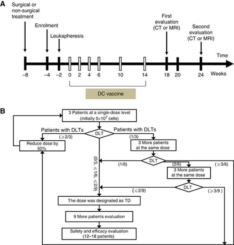
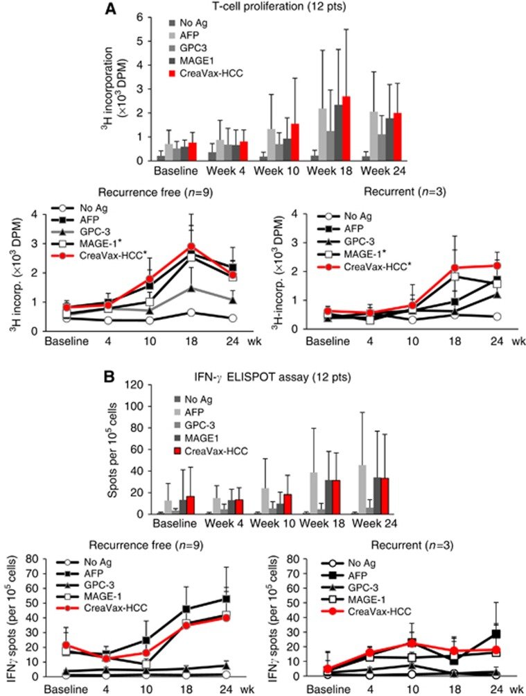
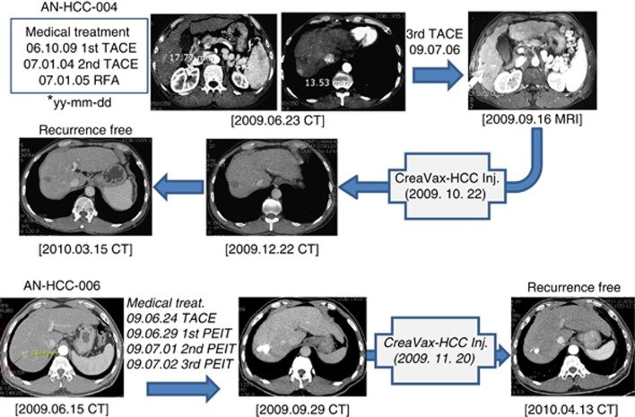
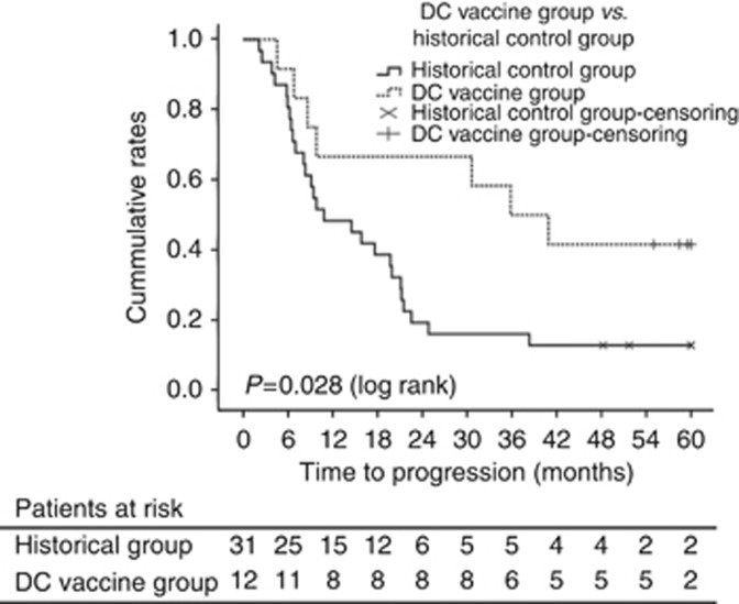
그림 출처: PubMed Central 공개 논문 (PMC4702003)
Conflict of interest statement
YL, MKHeo, and J-SS are CreaGene Inc. employees. Y-SB has been a consultant for JW CreaGene Inc. All remaining authors have declared no conflicts of interest.
그 밖의 정보
- 출판 형태
- Clinical Trial, Phase I, Clinical Trial, Phase II, Journal Article, Research Support, Non-U.S. Gov't
- 저널
- British journal of cancer · eISSN 1532-1827
- 전자 출판
- 2015-12-10
- MeSH
- Aged · Antigens, Neoplasm / immunology · Carcinoma, Hepatocellular / therapy · Cell Transplantation* · Dendritic Cells / immunology · Female · Humans · Immunotherapy* · Liver Neoplasms / therapy · Male · Middle Aged · Treatment Outcome
- Substances
- Antigens, Neoplasm
- 참고문헌
- 31편