
Sci Rep. 2021;11(1):17584.
Ultra-high-resolution computed tomography shows changes in the lungs related with airway hyperresponsiveness in a murine asthma model.
제1저자 정재우, 오정석, 배보람 · 교신저자 강혜련, 이창현
소속 (10)
- Institute of Allergy and Clinical Immunology, Seoul National University Medical Research Center, Seoul, Korea.
- Department of Internal Medicine, Chung-Ang University College of Medicine, Seoul, Korea.
- Department of Radiology, Catholic University, Seoul St. Mary's Hospital, Seoul, Korea.
- Department of Internal Medicine, Seoul National University College of Medicine, 103 Daehak-ro, Jongno-gu, Seoul, 03080, Korea.
- Department of Internal Medicine, University of Kansas School of Medicine, Kansas City, KS, USA.
- Department of Bioengineering, University of Kansas, Lawrence, KS, USA.
- Laboratory of Mucosal Immunology, Department of Biomedical Sciences, Seoul National University College of Medicine, Seoul, Korea.
- Institute of Allergy and Clinical Immunology, Seoul National University Medical Research Center, Seoul, Korea. helenmed@snu.ac.kr.
- Department of Internal Medicine, Seoul National University College of Medicine, 103 Daehak-ro, Jongno-gu, Seoul, 03080, Korea. helenmed@snu.ac.kr.
- Department of Radiology and Institute of Radiation, Seoul National University College of Medicine, Seoul National University Hospital, 103 Daehak-ro, Jongno-gu, Seoul, 03080, Korea. changhyun.lee@snu.ac.kr.
Abstract
In vivo presentation of airway hyper-responsiveness (AHR) at the different time points of the allergic reaction is not clearly understood. The purpose of this study was to investigate how AHR manifests in the airway and the lung parenchyma in vivo following exposure to different stimuli and in the early and late phases of asthma after allergen exposure. Ovalbumin (OVA)-induced allergic asthma model was established using 6-week female BALB/c mice. Enhanced pause was measured with a non-invasive method to assess AHR. The dynamic changes of the airway and lung parenchyma were evaluated with ultra-high-resolution computed tomography (128 multi-detector, 1024 × 1024 matrix) for 10 h. While the methacholine challenge showed no grossly visible changes in the proximal airway and lung parenchyma despite provoking AHR, the OVA challenge induced significant immediate changes manifesting as peribronchial ground glass opacities, consolidations, air-trapping, and paradoxical proximal airway dilatations. After resolution of immediate response, multiple episodes of AHRs occurred with paradoxical proximal airway dilatation and peripheral air-trapping in late phase over a prolonged time period in vivo. Understanding of airflow limitation based on the structural changes of asthmatic airway would be helpful to make an appropriate drug delivery strategy for the treatment of asthma.
© 2021. The Author(s).
Figures
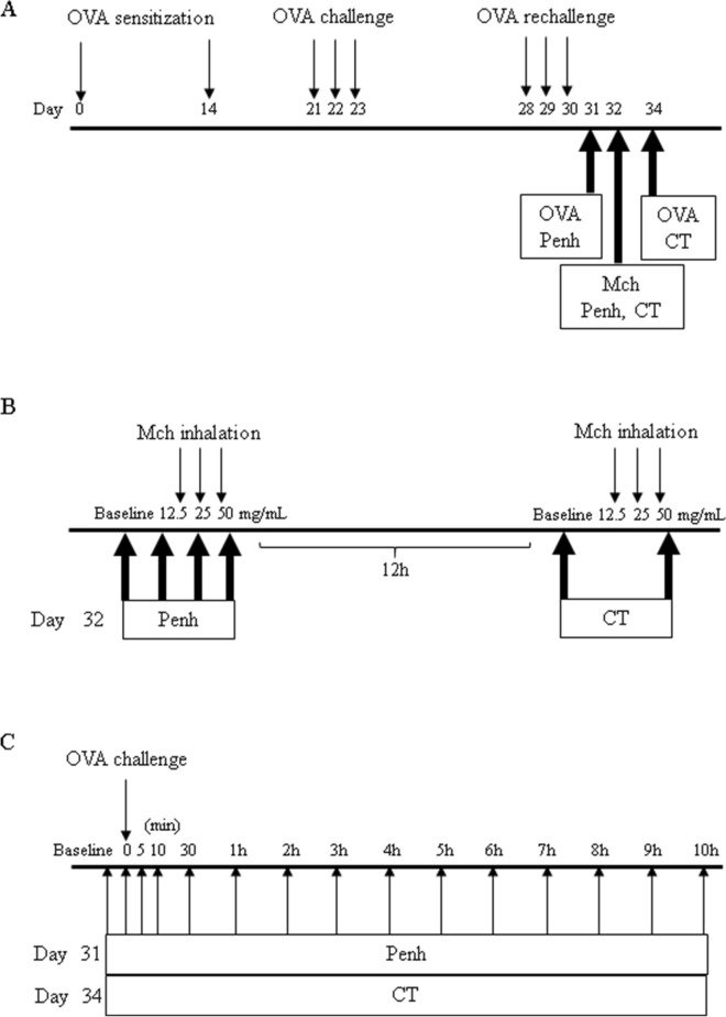
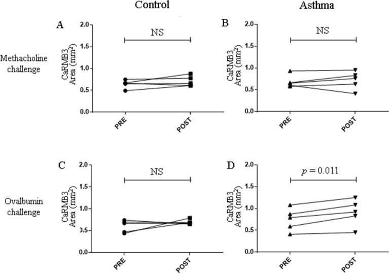
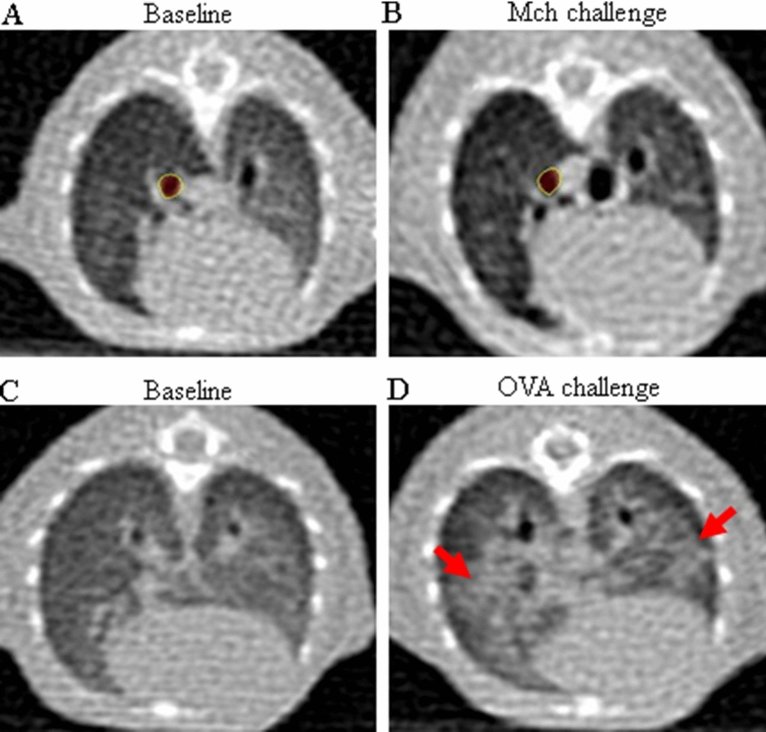
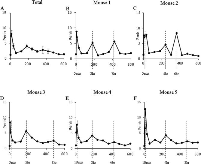
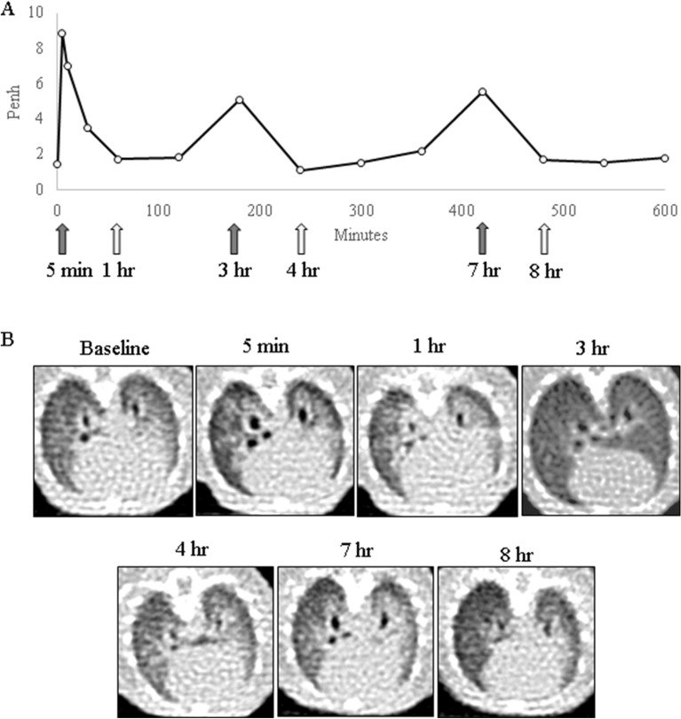
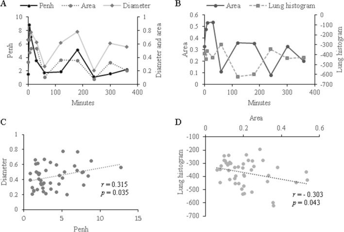
그림 출처: PubMed Central 공개 논문 (PMC8413288)
Conflict of interest statement
The authors declare no competing interests.
그 밖의 정보
- 출판 형태
- Journal Article, Research Support, Non-U.S. Gov't
- 저널
- Scientific reports · eISSN 2045-2322
- 전자 출판
- 2021-09-02
- MeSH
- Animals · Asthma / chemically induced / diagnostic imaging / pathology · Bronchial Hyperreactivity / diagnostic imaging / etiology / pathology · Disease Models, Animal · Female · Lung / diagnostic imaging / pathology · Mice · Mice, Inbred BALB C · Respiratory Hypersensitivity / diagnostic imaging / etiology / pathology · Tomography, X-Ray Computed
- 참고문헌
- 36편