College of Pharmacy, Research Institute of Pharmaceutical Sciences and Natural Products Research Institute, Seoul National University, Seoul, Republic of Korea.
Institute of Well-Aging Medicare & Chosun University LAMP Center, Chosun University, Gwangju, Republic of Korea.
Department of Pathology, University of Illinois Chicago, Chicago, IL, USA.
Department of Biomedical Sciences, Seoul National University College of Medicine, Seoul, Republic of Korea.
College of Pharmacy, Kangwon National University, Chuncheon, Republic of Korea.
Division of Endocrinology and Metabolism, Department of Internal Medicine, College of Medicine, Seoul Metropolitan Government Boramae Medical Center, Seoul National University, Seoul, Republic of Korea.
Division of Gastroenterology and Hepatology, Department of Internal Medicine, College of Medicine, Seoul Metropolitan Government Boramae Medical Center, Seoul National University, Seoul, Republic of Korea.
Department of Pathology, College of Medicine, Chosun University, Gwangju, Republic of Korea.
College of Pharmacy, Gachon University, Incheon, Republic of Korea.
Department of Biomedical Sciences, Seoul National University College of Medicine, Seoul, Republic of Korea. murimchoi@snu.ac.kr.
Division of Gastroenterology and Hepatology, Department of Internal Medicine, College of Medicine, Seoul Metropolitan Government Boramae Medical Center, Seoul National University, Seoul, Republic of Korea. drwon1@snu.ac.kr.
College of Pharmacy, Research Institute of Pharmaceutical Sciences and Natural Products Research Institute, Seoul National University, Seoul, Republic of Korea. kwkang@snu.ac.kr.
Abstract
A disintegrin and metalloproteinase with thrombospondin motifs 4 (ADAMTS4) has been implicated in arthritis and lung fibroblast activation; however, its role in liver homeostasis and fibrogenesis remains largely unexplored. Here, we investigated the functional significance of ADAMTS4 in liver fibrosis. We found that hepatic ADAMTS4 mRNA expression was significantly elevated in patients with fibrotic steatohepatitis. In mouse models of liver fibrosis, genetic deletion of ADAMTS4 protected against liver fibrogenesis, accompanied by a marked reduction in the recruitment of myeloid-derived infiltrating macrophages. Mechanistically, ADAMTS4-mediated cleavage of versican generated versikine, which promoted macrophage migration and differentiation toward a pro-inflammatory phenotype in vitro. In addition, tumor necrosis factor (TNF)α significantly increased both the mRNA expression and protein secretion of ADAMTS4. Furthermore, ADAMTS4 directly induced collagen accumulation through activation of signal transducer and activator of transcription 3 (STAT3) in LX2 cells. To explore the potential genetic regulation of ADAMTS4 expression, we performed response-eQTL analysis in patients with metabolic dysfunction-associated steatotic liver disease and identified a single-nucleotide polymorphism associated with increased ADAMTS4 expression in a subset of patients carrying a specific genotype. Collectively, our findings identify ADAMTS4 as a critical regulatory factor that promotes the recruitment of myeloid-derived infiltrating macrophages and collagen accumulation during liver fibrogenesis, suggesting that targeting ADAMTS4 may represent a potential therapeutic strategy for liver fibrosis.
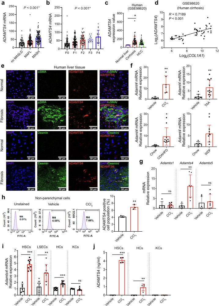
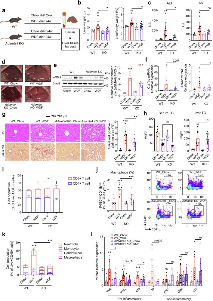
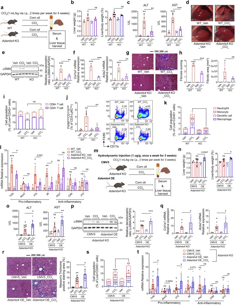
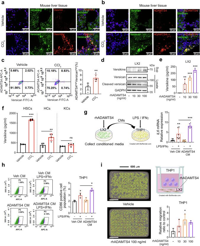
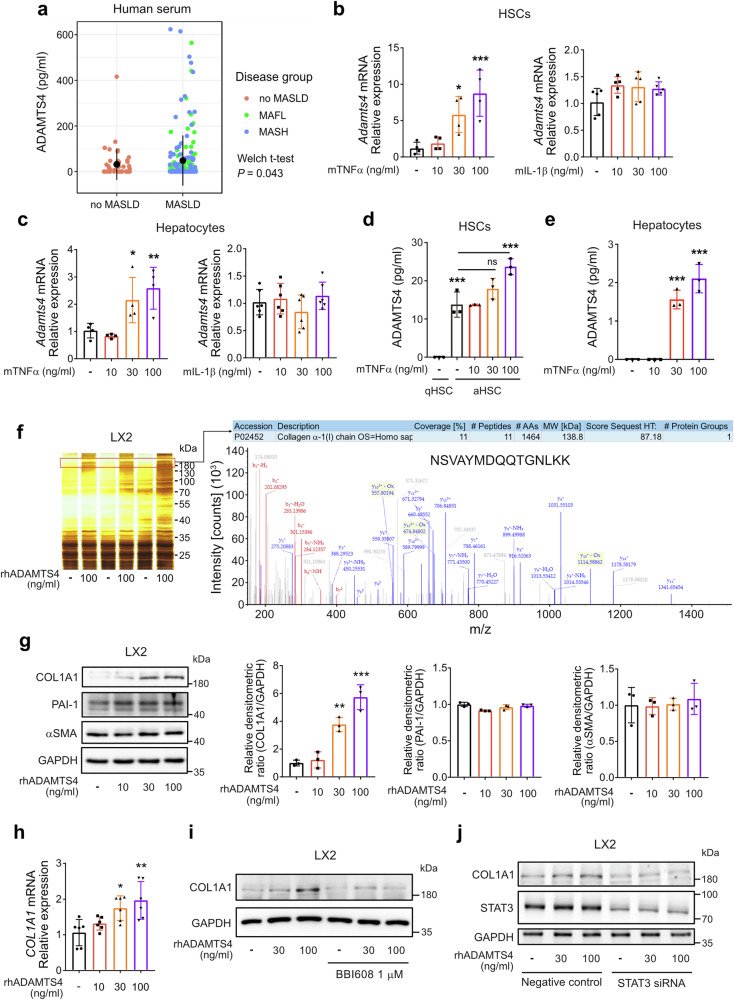
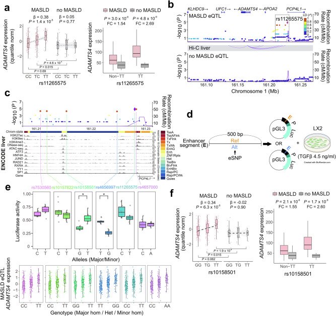
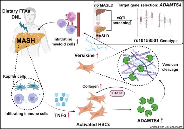
Fig. 1ADAMTS4 is upregulated in liver fibrosis in both humans and mice. a Hepatic ADAMTS4 mRNA expression according to the histological spectra of MASLD and b fibrosis severity in a biopsy-proven MASLD cohort (n = 293). The P value for trend was calculated using a generalized linear model. cADAMTS4 mRNA levels in normal, cirrhotic, and HCC livers from GEO data (GSE98620). d The correlation between hepatic COL1A1 and ADAMTS4 mRNA expression in GEO data (GSE98620) was analyzed using Pearson correlation analysis. e Immunofluorescence staining analysis of αSMA, ADAMTS4, and desmin expression in liver tissues from patients with fibrotic MASH. f mRNA levels of Adamts4 in liver tissues from chemical- or diet-induced mouse liver fibrosis models (n = 6–12). g mRNA levels of Adamts1, Adamts4 and Adamts5 in liver tissues from the CCl4-induced liver fibrosis model (n = 5–6). h Representative images (left) and cell population (right) of flow cytometric analyses of ADAMTS4 in non-parenchymal cells from mice 3 weeks after corn oil (vehicle) or CCl4 injection(n = 3). iAdamts4 mRNA levels in primary hepatocytes (HCs), liver sinusoidal endothelial cells (LSECs), hepatic stellate cells (HSCs), and Kupffer cells (KCs) from mice 3 weeks after vehicle or CCl4 injection (n = 8–12). j ADAMTS4 levels determined by ELISA in HCs, HSCs, and KCs from mice 3 weeks after vehicle or CCl4 injection (n = 4). Data are presented as mean ± SD. * indicates p ≤ 0.05, **p ≤ 0.01, ***p ≤ 0.001. MAFL metabolic dysfunction-associated fatty liverFig. 2Adamts4 deficiency suppresses myeloid-derived immune cells infiltration and stellate cell activation in WDF-induced liver fibrosis. a Schematic overview of the experimental setup. b Liver weight and liver-to-body weight ratio from C57BL/6 J WT and Adamts4 KO mice fed a normal chow diet or WDF for 24 weeks (n = 6–8). c Serum ALT and AST activities (n = 6–8). d Appearance of representative livers (n = 6–8). e) Immunoblotting analysis of αSMA and GAPDH expression in mouse liver tissues and densitometric analysis (n = 6–8). f mRNA levels of Col1a1 and Acta2. g Representative images of hematoxylin-and-eosin staining and Sirius Red staining of liver tissues. h Serum and liver TG levels (n = 6–8). i CD4+ T cell and CD8+ T cell populations were analyzed by flow cytometry. j Quantification graph of macrophage populations and representative images of flow cytometry of macrophages (F4/80high and CD11bhigh) (n = 6–8). k Neutrophil, monocyte, dendritic cell, and macrophage populations. l Relative mRNA expression of inflammatory cytokine genes (n = 6–8). Data are presented as mean ± SD. * indicates p ≤ 0.05, **p ≤ 0.01, ***p ≤ 0.001. Nos2 nitric oxide synthase-2, Arg1 arginase 1, Ciita class II transactivatorFig. 3Adamts4 deficiency suppresses myeloid-derived immune cells infiltration and stellate cell activation in CCl4-induced liver fibrosis. a Schematic overview of the experimental setup. b Liver weight and liver-to-body weight ratio from WT and Adamts4 KO mice after vehicle or CCl4 injection for 4 weeks (n = 3–4). c Serum ALT and AST activities (n = 3–4). d Representative livers (n = 3–4). e Immunoblotting analysis of αSMA and GAPDH (n = 3–4). f mRNA levels of Col1a1 and Acta2. g Representative images of Masson’s trichrome staining. h Fibrosis score and fibrotic area (n = 3–4). i CD4+ T cell and CD8+ T cell populations. j Quantification graph of macrophage populations and representative images of flow cytometry analysis (n = 3–4). k Neutrophil, monocyte, dendritic cell, and macrophage populations. l Relative mRNA expression of inflammatory cytokine genes (n = 3–4). m Schematic overview of the experimental setup. n Liver weight and liver-to-body weight ratio from control vector-injected and Adamts4 OE vector-injected Adamts4 KO mice after vehicle or CCl4 injection for 3 weeks (n = 4–8). o Serum ALT and AST activities (n = 4–8). p Immunoblotting analysis of αSMA and GAPDH (n = 4–8). q mRNA levels of Col1a1 and Acta2 (n = 4-8). r Representative images of Masson’s trichrome staining and quantification. s Neutrophil, monocyte, dendritic cell, and macrophage populations. t Relative mRNA expression of inflammatory cytokine genes (n = 4–8). Data are presented as mean ± SD * indicates p ≤ 0.05, **p ≤ 0.01, ***p ≤ 0.001. Nos2 nitric oxide synthase-2, Arg1 arginase 1, Ciita, class II transactivatorFig. 4ADAMTS4-derived versikine mediates pro-inflammatory macrophage recruitment. a Immunofluorescence staining of versican co-localization (yellow) with a stellate cell marker, αSMA. b ADAMTS4 co-localization with versican in mice after vehicle or CCl4 injection for 3 weeks. c Representative images of flow cytometry analysis of ADAMTS4 and versican in liver tissues from CCl4-injected mice for 3 weeks (n = 6). d Immunoblotting analysis of versican, cleaved versican, versikine, and GAPDH in LX2 cells treated with rhADAMTS4 for 24 h. e Versikine ELISA (n = 3). f Versikine levels determined by ELISA in primary hepatocytes (HCs), hepatic stellate cells (HSCs), and Kupffer cells (KCs) from mice 3 weeks after vehicle or CCl4 injection (n = 4). g Schematic overview of the experimental setup and mRNA level of IL6 in THP1 cells treated with 1 μg/ml LPS and 20 ng/ml IFNγ, together with the indicated conditioned media from LX2 cells for 24 h. h Representative images of flow cytometry analysis of THP1 cells to detect CD86 (n = 3). i THP1 cell migration in LX2 cells co-culture condition (n = 3), (red dots: migrated THP1 cells, yellow dots: confluency of THP1 cells, green dots: migration pores). Data are presented as mean ± SD. * indicates p ≤ 0.05, **p ≤ 0.01, ***p ≤ 0.001Fig. 5TNFα induces ADAMTS4 secretion in HSCs and hepatocytes, and secreted ADAMTS4 enhances COL1A1 expression. a ADAMTS4 ELISA in sera with no MASLD (n = 45), MAFL (n = 105), and MASH patients (n = 97). p-values were analyzed by the Welch t-test. bAdamts4 mRNA levels in HSCs treated with mouse TNFα or IL-1β for 20 h. cAdamts4 mRNA in hepatocytes treated with TNFα or IL-1β for 20 h (n = 4 or 6). d, e ADAMTS4 ELISA (n = 3). f Increased protein bands above 180 kDa in LX2 cells treated with rhADAMTS4 for 24 h. g Immunoblotting analysis of COL1A1, PAI-1, αSMA, and GAPDH in LX2 cells treated with rhADAMTS4 for 24 h and densitometric analysis results. h mRNA levels of COL1A1 in LX2 cells treated with rhADAMTS4 for 20 h. i Immunoblotting analysis of COL1A1 and GAPDH in LX2 cells treated with rhADAMTS4 for 24 h, with or without pretreatment with BBI608. j Effect of STAT3 siRNA on COL1A1 protein expression. Data are presented as mean ± SD. * indicates p ≤ 0.05, **p ≤ 0.01, ***p ≤ 0.001. q quiescent, a activated, MAFL metabolic dysfunction-associated fatty liverFig. 6Genotype-dependent expression of ADAMTS4 in MASLD. aADAMTS4 expression levels stratified by rs11265575 genotype and disease status. P values for genotype trends (β) were calculated using a generalized linear model, and pairwise comparisons were performed using two-tailed tests. b Genomic locations of six eQTL SNPs in a single linkage disequilibrium block and ADAMTS4 locus. c An enlarged view of the eQTL SNPs as indicated by the square in (b) and relevant ChIP-seq and ATAC-seq peaks. d Schematic diagram displaying the reporter analysis shown in (e) Allele-specific reporter gene analysis results of the six SNPs in LX2 cells treated with TGFβ. fADAMTS4 expression levels stratified by rs10158501 genotype and disease status. The right panel shows the expression of ADAMTS4 in a genotype-dependent manner. P values for genotype trends (β) were calculated using a generalized linear model, and pairwise comparisons were performed using two-tailed t tests. Data are presented as mean ± SD. * indicates Padj ≤ 0.05Fig. 7Proposed scheme of ADAMTS4-mediated immune cell recruitment and liver fibrogenesis in MASLD. In MASH, TNFα stimulates the expression and release of ADAMTS4 in hepatic stellate cells (HSCs). The released ADAMTS4 directly activates HSCs and cleaves versican to generate versikine, which promotes macrophage infiltration and contributes to liver fibrogenesis
그림 출처: PubMed Central 공개 논문 (PMC13149992)
Conflict of interest statement
Competing interests: The authors declare no competing interests. Ethics: All animals received humane care throughout the study. All animal protocols were approved by the IACUC at Seoul National University (SNU-220114-1-2, SNU-201102-3-3, and SNU-200720-3-2) and conducted in accordance with the criteria outlined in the Guide for the Care and Use of Laboratory Animals. For human liver samples, the study was conducted in accordance with the provisions of the Declaration of Helsinki for the participation of human subjects in research and was approved by the Institutional Review Board of Boramae Medical Center (IRB No. 16-2013-45). Written informed consent was obtained from each subject in the study cohort.
그 밖의 정보
출판 형태
Journal Article
저널
Signal transduction and targeted therapy · eISSN 2059-3635
ADAMTS4 Protein · STAT3 Transcription Factor · Adamts4 protein, mouse · ADAMTS4 protein, human
연구비
National Research Foundation of Korea (NRF) RS-2022-NR067269 · National Research Foundation of Korea (NRF) 2021M3A9E4021818 · National Research Foundation of Korea (NRF) RS-2022-NR067269 · National Research Foundation of Korea (NRF) 2021M3A9E4021818