Enhanced Generation of Human Induced Pluripotent Stem Cells from Peripheral Blood and Using Their Mesoderm Differentiation Ability to Regenerate Infarcted Myocardium.
New Drug Development Division, K-BioCELF Inc., Suwon, Gyeonggi-do, Republic of Korea.
Cardiovascular Center and Department of Internal Medicine, Seoul National University Hospital, Seoul, Republic of Korea.
Department of Convergence Medicine, Pusan National University School of Medicine, Yangsan, Gyeongsangnam-do, Republic of Korea.
Abstract
Тhe most pressing issue in generating induced pluripotent stem cells (iPSCs) in clinical practice is the cell source. Compared to human dermal fibroblasts (HDFs), which have been widely used, human peripheral blood could be a more easily obtainable alternative. However, iPSCs generated from fresh peripheral blood require inconvenient specific methods including isolation. Recently, we succeeded in isolating and culturing human heart-derived circulating cells called circulating multipotent stem (CiMS) cells. Here, we investigated the generation efficiency of CiMS-derived iPSCs (CiMS-iPSCs) and tested their differentiation potential into mesodermal lineages and cardiovascular cells. We isolated and cultured CiMS cells from peripheral mononuclear cells with a high efficiency. Moreover, our method succeeded in reprogramming the CiMS cells and generating iPSCs with higher efficiency compared to when HDFs were used. Compared to HDF-iPSCs or human embryonic stem cells (hESCs), CiMS-iPSCs showed high differentiation potential into mesodermal lineage cells and subsequently into endothelial cells, vascular smooth muscle cells, and cardiomyocytes. Further, we checked the epigenetic status of each cell type. While methylation of the CpG site of the brachyury T promoter did not differ between cell types, the histone H3 lysine 4 trimethylation level in the brachyury T promoter region was enhanced in CiMS-iPSCs, compared to that in other cell types. In contrast, histone H3 lysine 9 acetylation was downregulated during the differentiation process of the CiMS-iPSCs. In the myocardial infarction model, the CiMS-iPSCs group showed more therapeutic potential in regenerating the myocardium than other cell types. Our study showed a new method to isolate human heart-derived stem cells from human peripheral blood and to generate iPSCs efficiently. Due to epigenetic memory, these CiMS-iPSCs easily differentiated into cardiovascular lineage cells, resulting in improved efficiency in vivo. These results suggest that our new method using CiMS cells has therapeutic potential in regenerative medicine using cell therapy.
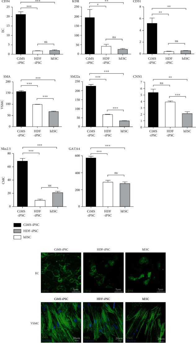
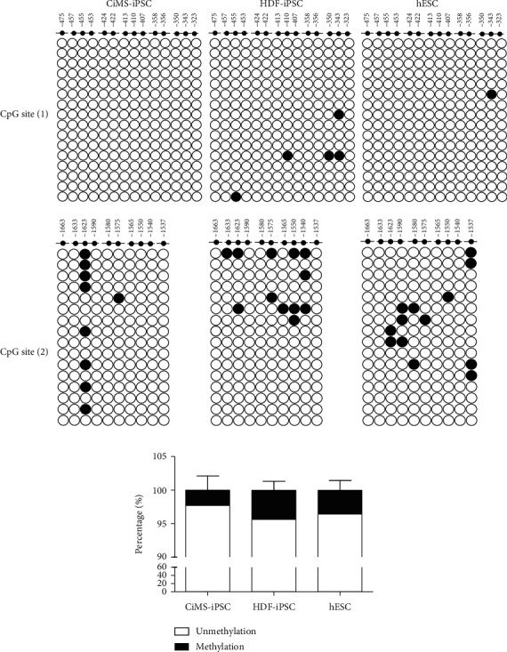
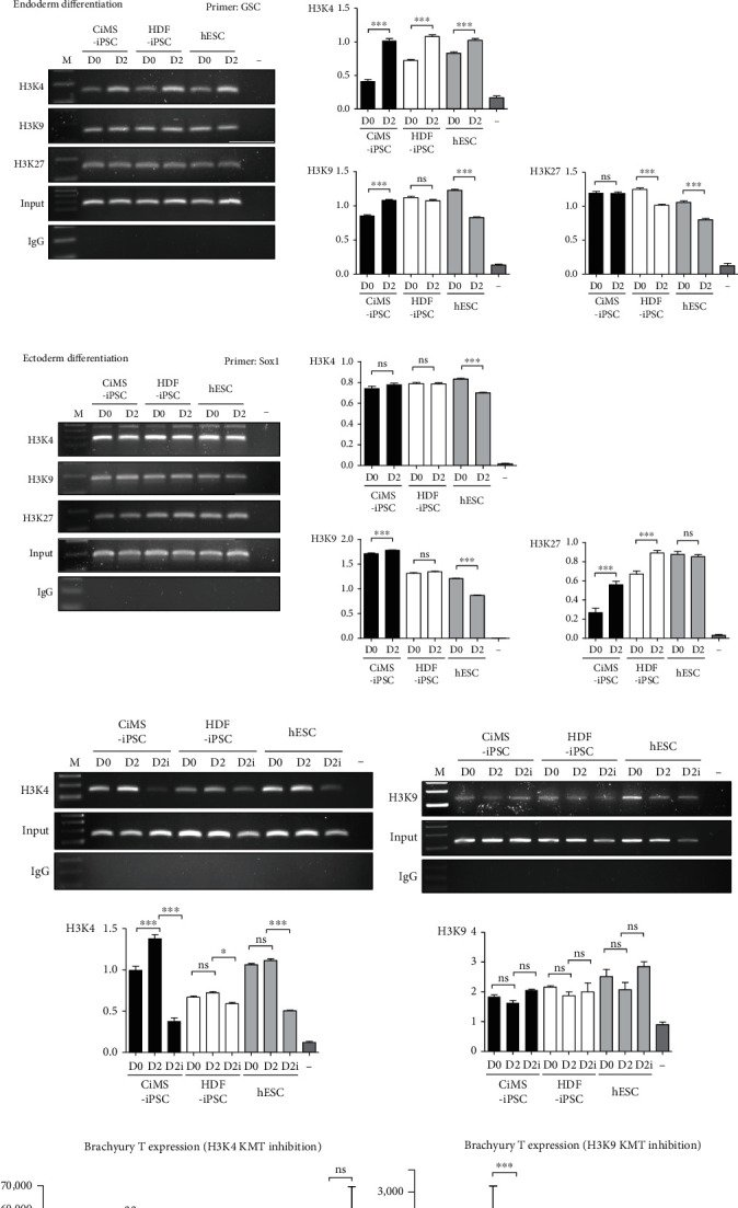
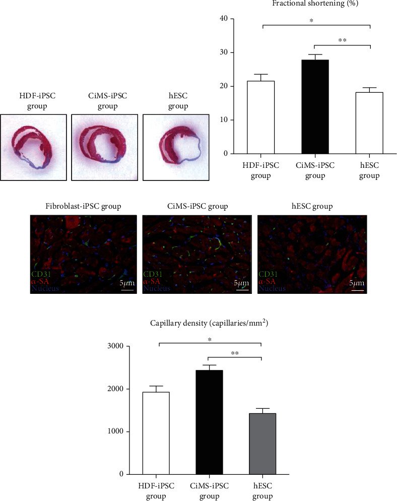
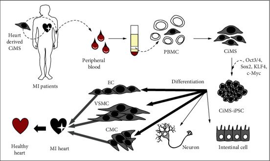
Figure 1Generation of induced pluripotent stem cells (iPSCs) from human circulating multipotent adult stem (CiMS) cells. (a) Time schedule of CiMS-iPSC generation. (b) CiMS morphology before reprogramming factor transduction and human embryonic stem cell- (hESC-) like CiMS-iPSC colonies on feeder cells after transduction. (c) CiMS-iPSCs were positive for ALP staining and expressed pluripotency markers, such as OCT3/4, NANOG, and TRA-1-81. (d) Transduction efficiency was estimated using GFP retroviral transduction. Retroviral transduction efficiency was higher in CiMS than in human dermal fibroblasts (HDFs) and even 293T cells (N = 3) (∗p < 0.05, ∗∗p < 0.01, and ∗∗∗p < 0.001: statistically significant, ns: statistically not significant). (e) Comparison of ALP-positive colony formation efficiency of CiMS cells and HDF (N = 4) (f) Expression levels of CiMS-iPSC pluripotency genes were similar to those in hESCs, confirmed by reverse transcription PCR. (g) The global gene expression profiles compared CiMS, CiMS-iPSCs, and hESCs (H9) with the oligonucleotide microarray. (h) Bisulfite genomic sequencing of the promoter regions of OCT3/4 and NANOG. Open and closed circles indicate unmethylated and methylated CpGs. (i) Karyotyping analyses of the CiMS-iPSCs. (j) CiMS-iPSCs were spontaneously differentiated into three germ layers, and immunofluorescence staining showed positivity for each marker. The markers used were beta III tubulin and nestin for ectoderm, alpha-sarcomeric actin and smooth muscle actin for mesoderm, and AFP for endoderm. (k) Teratoma derived from CiMS-iPSCs. H&E staining of teratoma derived from CiMS-iPSCs. CiMS-iPSCs were transplanted subcutaneously on the back of a SCID mouse.Figure 2Mesoderm differentiation preference in the process of CiMS-iPSC differentiation to three germ layers. (a) Expression of pluripotent markers on day 0 and day 2 of mesodermal differentiation was estimated by real-time PCR. The expression of early mesodermal markers on day 2 of mesoderm differentiation was compared in each cell type using real-time PCR. The expression of early endodermal markers on day 4 of endodermal differentiation and early ectodermal markers on day 5 of ectodermal differentiation was compared in each cell type using real-time PCR (∗p < 0.05, ∗∗p < 0.01, and ∗∗∗p < 0.001; ss: statistically significant; ns: statistically not significant; black bar: CiMS-iPSC; white bar: HDF-iPSC; grey bar: hESC). (b) After differentiation into cardiovascular lineage cells, the expression of endothelial cell (EC) markers (CD34, KDR, and CD31) on day 5 of EC differentiation, vascular smooth muscle cell (VSMC) markers (SMA, SM22a, and CNN1) on day 6 of VSMC differentiation, and cardiomyocyte (CMC) markers (Nkx2.5, GATA4) on day 12 of CMC differentiation was compared in each cell type using real-time PCR. (c) After differentiation into cardiovascular cells, such as ECs, VSMCs, and CMCs, cardiovascular cell-specific markers were detected by immunofluorescent staining, including CD31 for ECs, SMA for VSMCs, and alpha-sarcomeric actin for CMCs. (d) Marker-positive cells were calculated among the total differentiated cells, and differentiation efficiency was compared.Figure 3Comparison of brachyury T promoter methylation status in pluripotent cells. (a) Patterns of genomic DNA methylation at CpG site [1] (-475~-323) and CpG site [2] (-1537~1663) of brachyury T were confirmed by bisulfite PCR among the three different iPSCs. (b) Quantification of methylation status in the three cell types.Figure 4Comparison of histone tail modifications according to the three germline differentiation stages of each iPSC. (a) Histone tail modification levels of the transcription site of brachyury T during mesodermal differentiation (day 2 (D2)) of the three different cell types were compared using a chromatin immunoprecipitation (ChIP) assay. The H3K4me3, H3K9ac, and H3K27me3 levels of brachyury T were analyzed (∗p < 0.05, ∗∗p < 0.01, and ∗∗∗p < 0.001; ss: statistically significant; ns: statistically not significant). (b) Histone tail modification levels of the GSC transcription site during endodermal differentiation (day 2 (D2)) of the three different cell types were compared by ChIP assay. H3K4me3, H3K9ac, and H3K27me3 of GSC were analyzed. (c) Histone tail modification levels of the Sox1 transcription site during ectodermal differentiation (day 2 (D2)) of the three different cell types were compared by ChIP assay. H3K4me3, H3K9ac, and H3K27me3 of Sox1 were analyzed. (d) After treatment with the H3K4 KMT inhibitor (50 μM MM102; D1i, D2i) during mesodermal differentiation (day 1 (D1), day 2 (D2)), H3K4me3 level of the brachyury T transcription site was compared in each cell type. (e) After treatment with the H3K9 KMT inhibitor (100 nM chaetocin; D1i, D2i) during mesodermal differentiation (day 1 (D1), day 2 (D2)), the H3K9ac level of brachyury T transcription site was compared in each cell type. (f) Brachyury T gene expression following H3K4 KTM inhibitor (MM102) treatment on mesoderm differentiation day 1 and day 2 was measured in each cell type using real-time PCR. (g) Brachyury T gene expression following H3K4 KTM inhibitor (chaetocin) treatment on mesoderm differentiation day 1 and day 2 was measured in each cell type using real-time PCR.Figure 5In vivo potential of CiMS-iPSCs in myocardial infarction model. (a, b) To check the in vivo therapeutic potential of CiMS-iPSCs, we performed a myocardial infarction model in nude mice. The group injected with differentiated CiMS-iPSC showed significant improvement in infarct area reduction and cardiac systolic function (mean ± SEM, HDF-iPSC vs. CiMS-iPSC vs. hESCs: 21.57 ± 2.1% vs. 27.90 ± 1.56% vs. 18.21 ± 1.39%, p value < 0.001; n = 10 per group). (c) CiMS-iPSCs could differentiate into endothelial lineage cells more efficiently than other groups. (d) Quantitative data of capillary density in each group.Figure 6Production of iPSCs using CiMS cells and its application in cardiovascular cell differentiation. CiMS cells separated from the endocardium of the heart can be easily isolated from the blood and have high transduction efficiency; therefore, reprogramming is efficient. These CiMS cells are specialized for differentiation into cardiovascular cells even when they become iPSCs because of the epigenetic memory of the endocardium. Therefore, the generation of CiMS-iPSCs shows great advantages in cardiovascular disease-related modeling and is an appropriate cell line for stem cell therapy in regenerative medicine.
그림 출처: PubMed Central 공개 논문 (PMC8856835)
Conflict of interest statement
The authors declare that the research was conducted in the absence of any commercial or financial relationships that could be construed as a potential conflict of interest.