Department of Internal Medicine, Seoul National University College of Medicine, Seoul, Korea.
Department of Internal Medicine, Seoul National University College of Medicine, Seoul, Korea; Department of Molecular Medicine and Biopharmaceutical Sciences, Graduate School of Convergence Science and Technology, College of Medicine, Seoul National University, Seoul, Korea.
Abstract
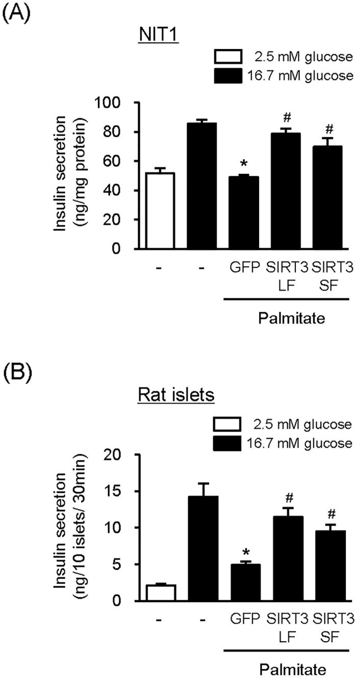
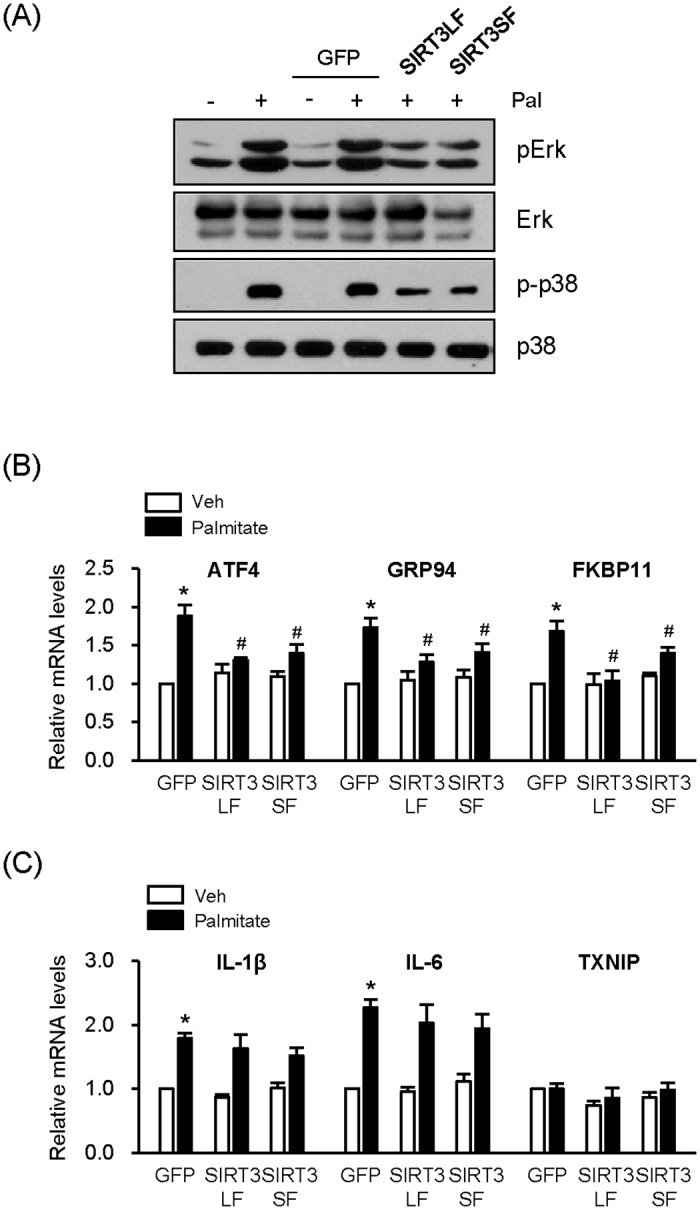
Abnormally high levels of circulating free fatty acids can lead to pancreatic islet β-cell dysfunction and apoptosis, contributing to β-cell failure in Type 2 diabetes. The NAD+-dependent protein deacetylase Sirtuin-3 (SIRT3) has been implicated in Type 2 diabetes. In this study, we tested whether SIRT3 overexpression affects palmitate-induced β-cell dysfunction in cells of line NIT1, which are derived from mouse pancreatic β-cells. Two different lengths of SIRT3 were overexpressed: full length SIRT3 (SIRT3LF), which was preferentially targeted to mitochondria and partially to the nucleus, and its N-terminal truncated form (SIRT3SF), which was located in the nucleus and cytoplasm. Overexpression of SIRT3LF and SIRT3SF using an adenoviral system alleviated palmitate-induced lipotoxicity such as reduction of cell viability and mitogen-activated protein kinase (MAPK) activation. Chronic exposure to low concentrations of palmitate suppressed glucose-stimulated insulin secretion, but the suppression was effectively reversed by overexpression of SIRT3LF or SIRT3SF. The mRNA levels of the endoplasmic reticulum (ER) stress responsive genes ATF4, GRP94 and FKBP11 were increased by palmitate treatment, but the increases were completely inhibited by SIRT3LF overexpression and less effectively inhibited by SIRT3SF overexpression. This result suggests that overexpression of SIRT3 inhibits induction of ER stress by palmitate. Collectively, we conclude that overexpression of SIRT3 alleviates palmitate-induced β-cell dysfunction.
Figures
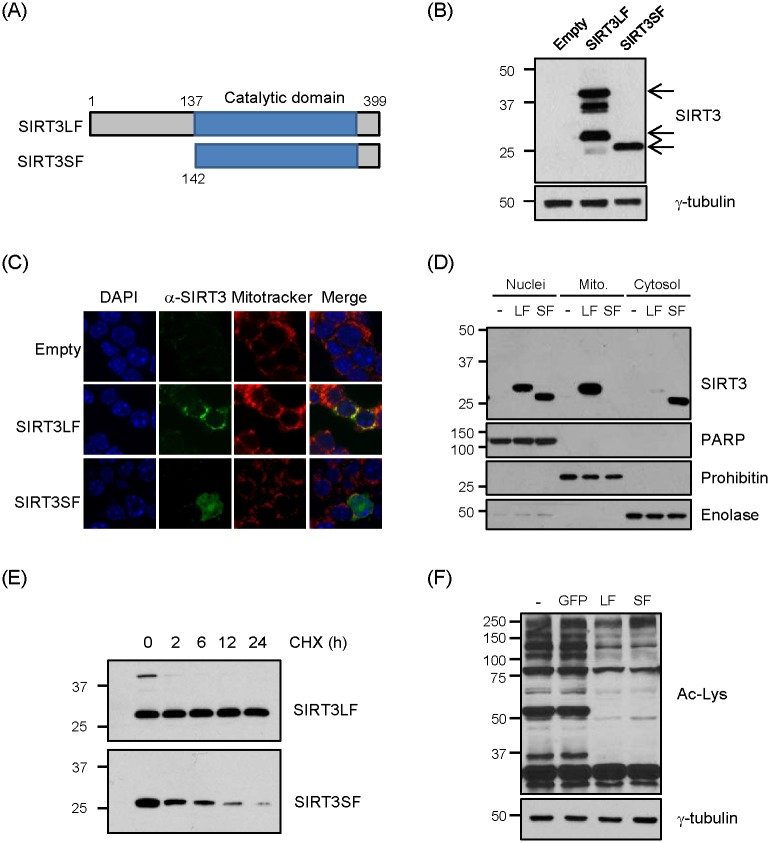
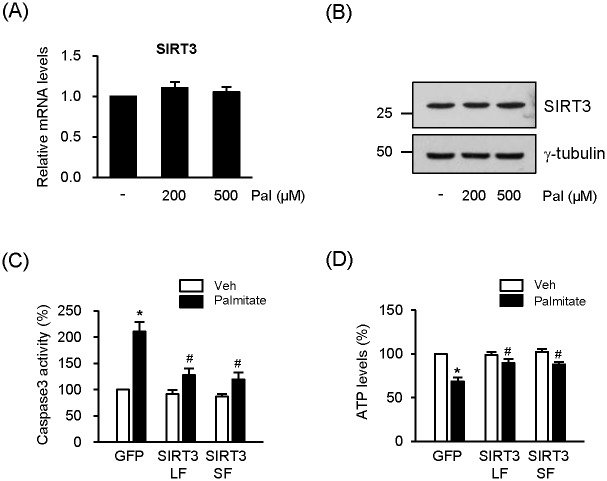
Fig 1 Localization of SIRT3LF and SIRT3SF. (A) Expression vectors and adenovirus expressing full-length human SIRT3 (SIRT3LF) and N-terminal region-truncated SIRT3 (SIRT3SF, with 141 N-terminal amino acids deleted) were generated. (B) NIT1 cells were infected with Ad-SIRT3LF (50 MOI), Ad-SIRT3SF (200 MOI) and Ad-β-gal (Empty, 200 MOI). Ten μg proteins were loaded on each lane and immunoblotted with a SIRT3 antibody. (C) NIT1 cells were infected with Ad-β-gal (Empty), Ad-SIRT3LF or Ad-SIRT3SF for 48 h. SIRT3 (green) was detected by immunofluorescence confocal microscopy. Mitochondria (red) were stained with Mitotracker, and nuclei were stained with DAPI (blue). (D) NIT1 cells were infected with Ad-SIRT3LF or Ad-SIRT3SF for 48h, and then subjected to subcellular fractionation. 15 μg proteins were used for Western blot analyses. Subcellular fractionation was confirmed by immunoblot with antibodies against PARP (nucleus), prohibitin (mitochondria) and enolase (cytosol). (E) COS7 cells were transfected with pcDNA-SIRT3LF (100 ng) or pcDNA-SIRT3SF (400 ng), and 24 h later the cells were treated with cycloheximide (CHX, 5 μM). Cell lysates were prepared after the CHX treatment, followed by Western blot analysis with anti-SIRT3 antibody. (F) NIT1 cells were infected with Ad-GFP (empty), Ad-SIRT3LF or Ad-SIRT3SF (all 50 MOI) for 48 h and trichostatin A (1 μM) was added 6 h before harvest. Whole cell lysates were subjected to Western blot analyses with anti-acetylated lysine (Ac-Lys) antibody.Fig 2 SIRT3 overexpression ameliorates reduction of viability by palmitate. (A) NIT1 cells were incubated with palmitate (Pal) as indicated. After 24 h incubation, SIRT3 mRNA levels were measured by real-time PCR. The mRNA level of untreated cells was expressed 1.0 and the others were as its relative values. Data are the means ± SEM of 5 independent experiments. (B) NIT1 cells were incubated with palmitate for 24h. Cell lysates (50 μg) were loaded on each lane of SDS-PAGE gels and immunoblotted with an anti-SIRT3 antibody. (C) NIT1 cells were infected with Ad-GFP, Ad-SIRT3LF, or Ad-SIRT3SF (all 50 MOI) and treated with palmitate (500 μM) for 24 h. Caspase 3 activity was measured [n = 6]. (D) NIT1 cells were infected with Ad-GFP, Ad-SIRT3LF, or Ad-SIRT3SF (all 50 MOI) and treated with palmitate (500 μM) for 8 h. Cellular ATP levels were determined [n = 4]. Caspase activity and ATP levels of the cells were expressed as percentage of values measured in the cells infected with Ad-GFP without palmitate. Data represent means ± SEM; *P < 0.05 for comparison with Ad-GFP infected cells without palmitate treatment; #P < 0.05 for the comparison with Ad-GFP infected cells treated with palmitate.Fig 3 SIRT3 overexpression partially recovers GSIS in the presence of palmitate. (A) NIT1 cells were infected with Ad-GFP, Ad-SIRT3LF, or Ad-SIRT3SF (50 MOI) for 24 h and then treated with palmitate (200 μM) for additional 48 h. Secreted insulin was measured after incubation with low glucose (2.5 mM) or high glucose (16.7 mM) for 1 h. Data represent as means ± SEM [n = 6]; *P < 0.05 for the comparison with the value of high glucose treated cells without virus infection or palmitate treatment; #P < 0.05 for the comparison with the value of cells infected with Ad-GFP and treated with palmitate. (B) Pancreatic islets isolated from rats were infected with Ad-GFP, Ad-SIRT3LF, or Ad-SIRT3SF (100 MOI) for 24h and incubated with palmitate (400 μM) for additional 48 h. Secreted insulin from 10 islets was measured 30 min after each glucose exposure. Data represent as means ± SEM [n = 3]; *P < 0.05 for the comparison with the value of high glucose treated cells without virus infection or palmitate treatment; #P < 0.05 for the comparison with the value of cells infected with Ad-GFP and treated with palmitate.Fig 4 SIRT3 overexpression inhibits palmitate-induced MAPK activation and ER stress. (A) NIT1 cells were infected with Ad-GFP, Ad-SIRT3LF, or Ad-SIRT3SF (50 MOI) and treated with palmitate (500 μM) for 8 h. The cell lysates were subjected to Western blot analysis. (B and C) NIT1 cells were infected with Ad-GFP, Ad-SIRT3LF, or Ad-SIRT3SF (50 MOI) and treated with palmitate (500 μM) for 24 h. The mRNA levels of ATF4, GRP94, and FKBP11 [n = 6] (B) and the mRNA levels of IL-1β, IL-6 and TXNIP [n = 5] (C) were measured using real-time PCR. The mRNA levels of the cells infected with Ad-GFP without palmitate treatment was set to 1.0 and the others were expressed relative to that value; *P < 0.05 for the comparison with the value of the Ad-GFP-infected cells without palmitate; #P < 0.05 for the comparison with the value of Ad-GFP infected cells in the presence of palmitate.
그림 출처: PubMed Central 공개 논문 (PMC4411148)
Conflict of interest statement
Competing Interests:The authors have declared that no competing interests exist.
그 밖의 정보
출판 형태
Journal Article, Research Support, Non-U.S. Gov't
저널
PloS one · eISSN 1932-6203
전자 출판
2015-04-27
MeSH
Adenosine Triphosphate · Animals · Caspase 3 / metabolism · Cell Death / drug effects / genetics · Cell Line · Diabetes Mellitus, Type 2 / genetics / metabolism · Endoplasmic Reticulum Stress / drug effects · Gene Expression* · Glucose / metabolism · Humans · Insulin / metabolism · Insulin-Secreting Cells / drug effects / metabolism · Islets of Langerhans / metabolism · MAP Kinase Signaling System / drug effects · Mice · Mice, Transgenic · Palmitates / pharmacology · Protein Transport · Rats · Sirtuin 3 / genetics / metabolism