
PLoS One. 2015;10(4):e0123094.
Thalidomide inhibits alternative activation of macrophages in vivo and in vitro: a potential mechanism of anti-asthmatic effect of thalidomide.
제1저자 이현승, 권혁수 · 교신저자 강혜련, 장윤석
소속 (5)
- Institute of Allergy and Clinical Immunology, Seoul National University Medical Research Center, Seoul, Korea.
- Institute of Allergy and Clinical Immunology, Seoul National University Medical Research Center, Seoul, Korea; Department of Internal medicine, Seoul National University College of Medicine, Seoul, Korea.
- Department of Medical Science, Seoul National University College of Medicine, Seoul, Korea.
- Department of Anatomy, Seoul National University College of Medicine, Seoul, Korea.
- Department of Internal Medicine, Seoul National University Bundang Hospital, Seongnam, Gyeonggi-do, Korea.
Abstract
BACKGROUND Thalidomide is known to have anti-inflammatory and immunomodulatory actions. However, the effect and the anti-asthmatic mechanism of thalidomide in the pathogenesis of asthmatic airways are not fully understood.
OBJECTIVE This study is designed to determine the effect and the potential mechanism of thalidomide in the pathogenesis of asthmatic airways using animal model of allergic asthma.
METHODS Six-week-old female BALB/C mice were sensitized with alum plus ovalbumin (OVA) and were exposed to OVA via intranasal route for 3 days for challenge. Thalidomide 200 mg/kg was given via gavage twice a day from a day before the challenge and airway hyperresponsivenss (AHR), airway inflammatory cells, and cytokines in bronchoalveolar lavage fluids (BALF) were evaluated. The expression levels of pro-inflammatory cytokines and other mediators were evaluated using ELISA, real time (RT)-qPCR, and flow cytometry. CRL-2456, alveolar macrophage cell line, was used to test the direct effect of thalidomide on the activation of macrophages in vitro.
RESULTS The mice with thalidomide treatment showed significantly reduced levels of allergen-induced BALF and lung inflammation, AHR, and the expression of a number of pro-inflammatory cytokines and mediators including Th2 related, IL-17 cytokines, and altered levels of allergen-specific IgG1/IgG2a. Of interesting note, thalidomide treatment significantly reduced expression levels of allergen- or Th2 cytokine-stimulated alternative activation of macrophages in vivo and in vitro.
CONCLUSION These studies highlight a potential use of thalidomide in the treatment of allergic diseases including asthma. This study further identified a novel inhibitory effect of thalidomide on alternative activation of macrophages as a potential mechanism of anti-asthmatic effect of thalidomide.
Figures


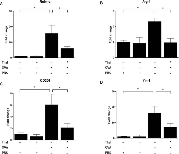


그림 출처: PubMed Central 공개 논문 (PMC4408009)
Conflict of interest statement
Competing Interests:The authors have declared that no competing interests exist.
그 밖의 정보
- 출판 형태
- Journal Article, Research Support, Non-U.S. Gov't
- 저널
- PloS one · eISSN 1932-6203
- 전자 출판
- 2015-04-23
- MeSH
- Allergens / pharmacology · Alum Compounds / pharmacology · Animals · Anti-Asthmatic Agents / pharmacology · Anti-Inflammatory Agents / pharmacology · Asthma / chemically induced / drug therapy / metabolism · Bronchial Hyperreactivity / chemically induced / drug therapy / metabolism · Bronchoalveolar Lavage Fluid · Disease Models, Animal · Female · Immunoglobulin G / metabolism · Inflammation / chemically induced / drug therapy / metabolism · Interleukin-17 / metabolism · Macrophages, Alveolar / drug effects / metabolism · Mice · Mice, Inbred BALB C · Ovalbumin / pharmacology · Th2 Cells / drug effects / metabolism · Thalidomide / pharmacology
- Substances
- Allergens · Alum Compounds · Anti-Asthmatic Agents · Anti-Inflammatory Agents · IL17A protein, human · Immunoglobulin G · Interleukin-17 · ovalbumin-alum · Thalidomide · Ovalbumin
- 참고문헌
- 42편