Department of Internal Medicine, Seoul National University Bundang Hospital, Bundang-gu, Seongnam-si, Gyeonggi-do, 13620, Republic of Korea.
Department of Internal Medicine, Seoul National University College of Medicine, Seoul, 03080, Republic of Korea.
Department of Internal Medicine, Seoul National University Bundang Hospital, Bundang-gu, Seongnam-si, Gyeonggi-do, 13620, Republic of Korea. kingfish12@naver.com.
Department of Internal Medicine, Seoul National University Bundang Hospital, Bundang-gu, Seongnam-si, Gyeonggi-do, 13620, Republic of Korea. woltoong@snu.ac.kr.
Department of Internal Medicine, Seoul National University College of Medicine, Seoul, 03080, Republic of Korea. woltoong@snu.ac.kr.
Abstract
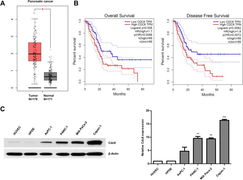
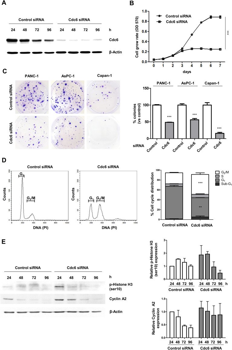
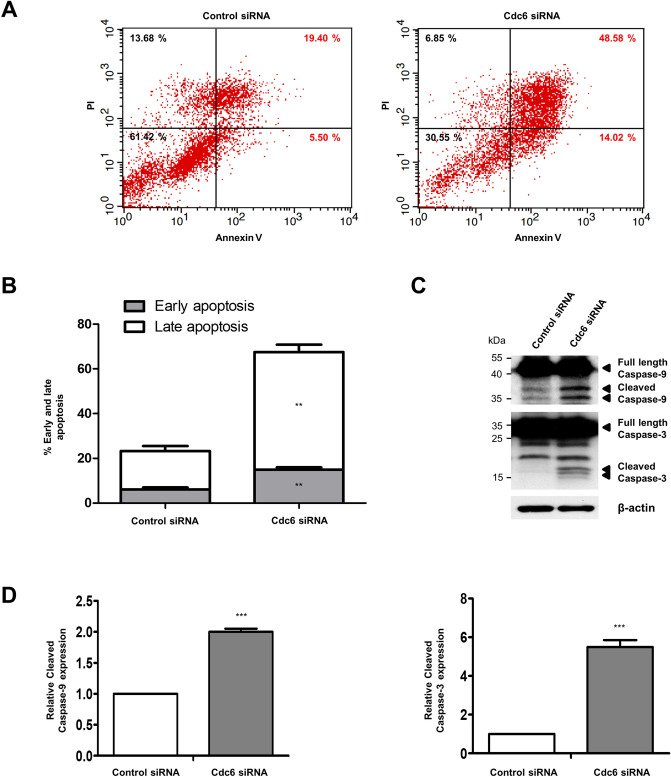
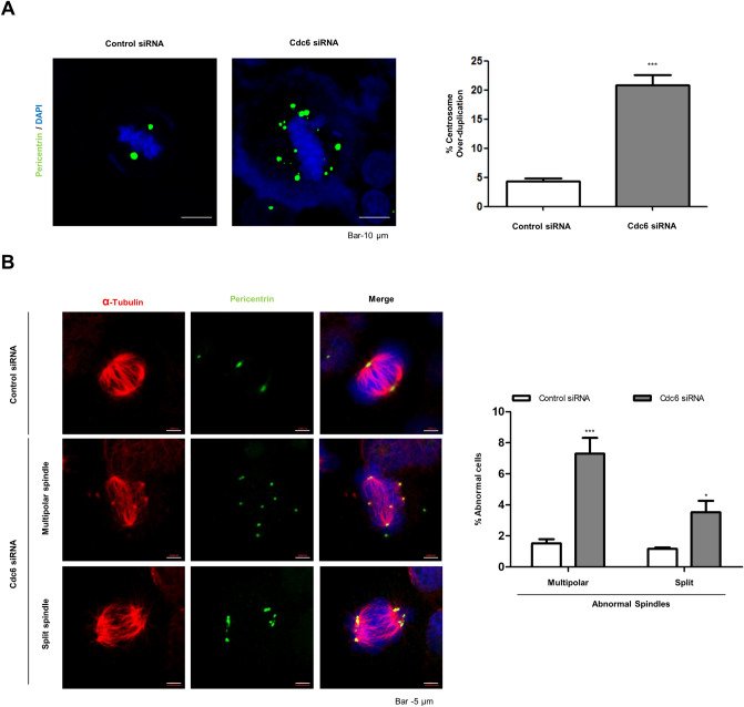
Cell division cycle 6 (Cdc6) plays key roles in regulating DNA replication, and activation and maintenance of cell cycle check points. In addition, Cdc6 exerts oncogenic properties via genomic instability associated with incomplete DNA replication. This study aimed to examine the effects of Cdc6 on pancreatic cancer (PC) cells. Our results showed that Cdc6 expression was higher in clinical PC specimens (based on analysis of the GEPIA database) and cell lines, and the high Cdc6 expression was associated with poorer survival in The Cancer Genome Atlas-PC cohort. In addition, Cdc6-depleted PC cells significantly inhibited cell proliferation and colony formation, delayed G2/M cell cycle progression, and increased expression of p-histone H3 and cyclin A2 levels. These observations could be explained by Cdc6 depletion leading to multipolar and split spindles via centrosome amplification and microtubule disorganization which eventually increases chromosome missegregation. Furthermore, Cdc6-depleted PC cells showed significantly increased apoptosis, which was consistent with increased caspase-9 and caspase-3 activation. Collectively, our results demonstrated that Cdc6-depleted PC cells are arrested in mitosis and eventually undergo cell death by induced multipolar spindles, centrosome aberrations, microtubule disorganization, and chromosome instability. In conclusion, Cdc6 may be a potential biomarker and therapeutic target for PC.
Figures
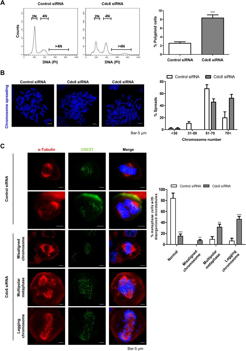
Figure 1Cdc6 is upregulated in PC and correlates with clinical effects. (A) Cdc6 mRNA expression levels in PC tissues (n = 179) and normal tissues (n = 171) based on the TCGA and GTEx database. Each dot represents Cdc6 expression in one sample. (B) The OS and DFS of PC patients with low or high Cdc6 expression levels were analyzed using the Kaplan–Meier method and a log rank test in PC. Median values are indicated by full lines. *P < 0.005. TCGA, The Cancer Genome Atlas; TPM, transcripts per million; OS, overall survival; DSF, disease free survival; HR, hazard ratio. (C) Cdc6 expression was detected in PC cell lines (AsPC-1, PANC-1, MIA PaCa-2 and Capan-1 cells), HUVEC, and HPDE pancreatic cells by western blotting. The Cdc6/β-actin ratio was determined by densitometric analysis using ImageJ. Error bars represent standard deviations of the means of three biological replicates. Statistical analysis was performed using t-test. **p < 0.001, ***p < 0.0001.Figure 2Cdc6 depletion suppresses PC cell proliferation and colony formation and induces G2/M cell cycle arrest. (A) PANC-1 cells were transfected with siRNA specific for Cdc6 or control. The level of Cdc6 proteins were confirmed by western blotting. Cells were harvested at the indicated time and lysates were immunoblotted with the indicated antibodies. (B) The effects of Cdc6 depletion on PANC-1 cell proliferation were determined using MTT assays. Cells were transfected with control and Cdc6 siRNA for 7 days. Error bars represent standard deviations of the means of three biological replicates. Statistical analysis was performed using two-way analysis of variance. ***p < 0.0001. (C) Colony formation assay was performed in PANC-1, AsPC-1 and Capan-1 cells with control siRNA and Cdc6 siRNA. The colonies were imaged and counted. Error bars represent standard deviations of the means of three biological replicates. Statistical analysis was performed using t-test. ***p < 0.0001. (D) Flow cytometric profiles of PANC-1 cells with control and Cdc6 siRNA for 72 h. Cells were harvested and analyzed to determine the percentage of cells in sub-G1, G1, S, and G2/M phase. Relative quantitative analysis data from a representative of three experiments are shown. Statistical analysis was performed using t-test. **p < 0.001, ***p < 0.0001. (E) Western blot analysis of the cell cycle-related proteins p-histone H3 and cyclin A2 in PANC-1 cells. Cells were harvested at the indicated time and lysates were immunoblotted with the indicated antibodies. The p-histone H3/β-actin and cyclin A2/β-actin ratios were determined by densitometric analysis using ImageJ.Figure 3Cdc6 depletion induces apoptosis in PC cells. (A) Apoptotic cells were detected by Annexin V-FITC and PI staining using flow cytometry of PANC-1 cells with control or Cdc6 siRNA. Early apoptotic cells stained as Annexin V-positive and PI-negative (lower right), and late apoptotic cells stained as Annexin V-positive and PI-positive (upper right). (B) The percentage of cells including early apoptotic cells and late apoptotic cells. Relative quantitative analysis data from a representative of three experiments are shown. Statistical analysis was performed using t-test. **p < 0.001. (C) Western blot analysis was performed to determine the expression of the apoptosis-related proteins caspase-9 and caspase-3. Cells were harvested at the indicated time and lysates were immunoblotted with the indicated antibodies. (D) The cleaved caspase-9/β-actin and cleaved caspase-3/β-actin ratios were determined by densitometric analysis using ImageJ. Error bars represent standard deviations of the means of three biological replicates. Statistical analysis was performed using t-test. ***P < 0.0001.Figure 4Cdc6 depletion causes centrosome abnormalities in PC cells. (A) Immunofluorescence staining of PANC-1 cells transfected with control or Cdc6 siRNA with pericentrin (green) antibodies and DAPI (blue). Graph represents the percentage of cells with centrosome over-duplication and chromosome abnormalities. Scale bar, 10 µm. Relative quantitative analysis data from a representative of three experiments are shown. Statistical analysis was performed using t-test. ***p < 0.0001. (B) Immunofluorescence staining of PANC-1 cells transfected with control or Cdc6 siRNA with α-tubulin (red), pericentrin (green) antibodies, and DAPI (blue). The graph represents the percentage of cells with abnormal spindles into multipolar and split spindles. Scale bar, 5 µm. Data are representative of three different fields in each independent experiment. Statistical analysis was performed using t-test. *P < 0.005, ***p < 0.0001.Figure 5Cdc6 depletion induces chromosome instability. (A) Flow cytometry of polyploidy cells (> 4 N DNA content) with control or Cdc6 siRNA in PANC-1 cells for 96 h. Graph represents the percentage of polyploidy cells. Statistical analysis was performed using t-test. ***p < 0.0001. (B) Aneuploidy cells were represented by metaphase chromosome spread analysis with control or Cdc6 siRNA in PANC-1 cells for 72 h. Spread chromosomes were observed by staining with DAPI and chromosome number divided aneuploidy into < 30, 31–50, 51–70, and > 70 chromosome numbers. Scale bar, 5 µm. (C) Immunofluorescence staining of PANC-1 cells transfected with control or Cdc6 siRNA with α-tubulin (red), CREST (green) antibodies, and DAPI (blue). The percentage of cells with misaligned chromosome, multipolar metaphase, and lagging chromosome was measured. Scale bar, 5 µm. Data are representative of three different fields were analyzed each independent experiment. Statistical analysis was performed using t-test. **p < 0.001, ***p < 0.0001.