
Sci Rep. 2020;10(1):19867.
Toll-like receptor 4 regulates intestinal fibrosis via cytokine expression and epithelial-mesenchymal transition.
제1저자 전유경 · 교신저자 고성준, 김지원
소속 (7)
- Department of Internal Medicine and Liver Research Institute, Seoul National University College of Medicine, 101 Daehak-ro, Jongno-gu, Seoul, 03080, Republic of Korea.
- Laboratory of Intestinal Mucosa and Skin Immunology, Seoul National University College of Medicine, 101 Daehak-ro, Jongno-gu, Seoul, 03080, Republic of Korea.
- Department of Dermatology, Boramae Medical Center, 20 Boramae-ro-5-gil, Dongjak-gu, Seoul, 07061, Republic of Korea.
- Division of Gastroenterology, Department of Internal Medicine, Boramae Medical Center, 20 Boramae-ro-5-gil, Dongjak-gu, Seoul, 07061, Republic of Korea. kjwjor@snu.ac.kr.
- Laboratory of Intestinal Mucosa and Skin Immunology, Seoul National University College of Medicine, 101 Daehak-ro, Jongno-gu, Seoul, 03080, Republic of Korea. kjwjor@snu.ac.kr.
- Department of Internal Medicine and Liver Research Institute, Seoul National University College of Medicine, 101 Daehak-ro, Jongno-gu, Seoul, 03080, Republic of Korea. jel1206@snu.ac.kr.
- Laboratory of Intestinal Mucosa and Skin Immunology, Seoul National University College of Medicine, 101 Daehak-ro, Jongno-gu, Seoul, 03080, Republic of Korea. jel1206@snu.ac.kr.
Abstract
Intestinal fibrosis induced by chronic and recurrent colitis, which is exacerbated by bowel stenosis, stricture, and obstruction, is challenging to treat. Toll-like receptor 4 (TLR4) stimulates innate and acquired immunity in response to specific microbial components, but the role of TLR4 in intestinal fibrosis is largely unknown. We investigated its role in intestinal fibrosis using not only a murine fibrosis model but also human myofibroblasts and intestinal epithelial cells. Colon fibrosis was induced in TLR4-deficient (TLR4-/-) mice and its wild-type counterparts with 3% dextran sulfate sodium. Absence of TLR4 gene attenuated chronic inflammation and colonic macrophages infiltration; intestinal fibrosis and collagen deposition were suppressed. Also, the production of tumor necrosis factor-α, interleukin-12p40, and transforming growth factor-β was reduced in TLR4-deficient peritoneal macrophages. TLR4 was silenced in CCD-18Co cells by small interfering RNA (siRNA), and matrix metalloproteinase-1, tissue inhibitor of metalloproteinase, and collagen α1 expression was evaluated. Role of TLR4 in epithelial-mesenchymal transition (EMT) was evaluated in HCT116 cells. Suppression of TLR4 transcription by siRNAs affected myofibroblasts activity, collagen synthesis, and EMT in the human cancer cell line. Thus, we suggest that TLR4 can be an essential mediator in intestinal chronic inflammation and fibrosis, indicating that TLR4 signaling is a potential therapeutic target for intestinal fibrosis.
Figures
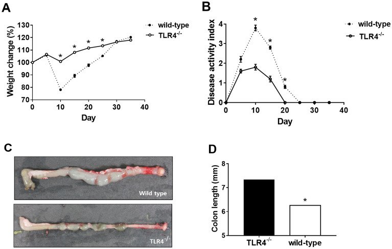
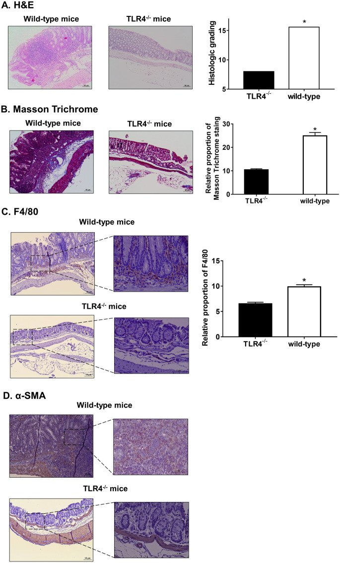
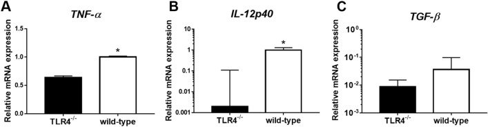
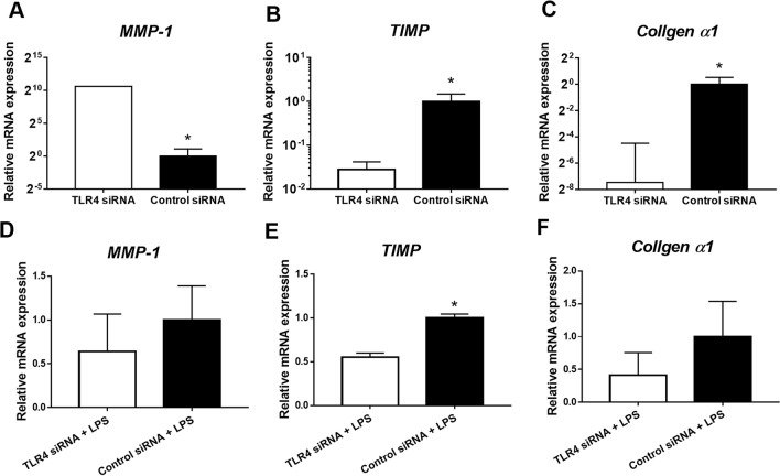
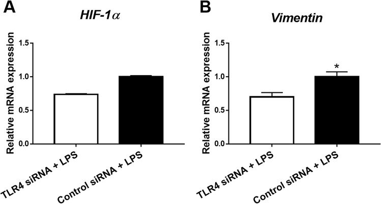
그림 출처: PubMed Central 공개 논문 (PMC7670421)
Conflict of interest statement
The authors declare no competing interests.
그 밖의 정보
- 출판 형태
- Journal Article, Research Support, Non-U.S. Gov't
- 저널
- Scientific reports · eISSN 2045-2322
- 전자 출판
- 2020-11-16
- MeSH
- Animals · Cell Line · Collagen · Colonic Diseases / chemically induced / genetics / immunology / pathology · Cytokines / metabolism · Dextran Sulfate / adverse effects · Disease Models, Animal · Epithelial-Mesenchymal Transition · Fibrosis · Gene Expression Regulation / drug effects · HCT116 Cells · Humans · Interleukin-12 Subunit p40 / metabolism · Macrophages, Peritoneal / metabolism · Mice · Signal Transduction · Toll-Like Receptor 4 / genetics · Transforming Growth Factor beta / metabolism · Tumor Necrosis Factor-alpha / metabolism
- Substances
- Cytokines · Interleukin-12 Subunit p40 · TLR4 protein, human · Tlr4 protein, mouse · Toll-Like Receptor 4 · Transforming Growth Factor beta · Tumor Necrosis Factor-alpha · Collagen · Dextran Sulfate
- 참고문헌
- 49편