
PLoS One. 2015;10(10):e0140421.
Intravascular Ultrasound and Angiographic Predictors of In-Stent Restenosis of Chronic Total Occlusion Lesions.
제1저자 강지훈 · 교신저자 조영석
소속 (3)
- Department of Internal Medicine, Seoul National University Bundang Hospital, Seongnam, South Korea; Department of Internal Medicine, Seoul National University Hospital, Seoul, South Korea.
- Department of Internal Medicine, Seoul National University Bundang Hospital, Seongnam, South Korea; Department of Internal Medicine, Seoul National University College of Medicine, Seoul, South Korea.
- Department of Internal Medicine, Seoul National University Bundang Hospital, Seongnam, South Korea.
Abstract
Despite the benefits of successful percutaneous coronary interventions (PCIs) for chronic total occlusion (CTO) lesions, PCIs of CTO lesions still carry a high rate of adverse events, including in-stent restenosis (ISR). Because previous reports have not specifically investigated the intravascular ultrasound (IVUS) predictors of ISR in CTO lesions, we focused on these predictors. We included 126 patients who underwent successful PCIs, using drug-eluting stents, and post-PCI IVUS of CTO lesions. Patient and lesion characteristics were analyzed to elucidate the ISR predictors. In each lesion, an average of 1.7 ± 0.7 (mean length, 46.4 ± 20.3 mm) stents were used. At 9 months follow-up, 14 (11%) patients demonstrated ISR, and 8 (6.3%) underwent target lesion revascularization. Multivariate logistic regression analysis showed that the independent predictors of ISR were the post-PCI minimal luminal diameter (MLD) and the stent expansion ratio (SER; minimal stent cross-sectional area (CSA) over the nominal CSA of the implanted stent), measured using quantitative coronary angiography (QCA) and IVUS, respectively. A receiver operating characteristic analysis indicated that the best post-PCI MLD and SER cut-off values for predicting ISR were 2.4 mm (area under the curve [AUC], 0.762; 95% confidence interval (CI), 0.639-0.885) and 70% (AUC, 0.714; 95% CI, 0.577-0.852), respectively. Lesions with post-PCI MLD and SER values less than these threshold values were at a higher risk of ISR, with an odds ratio of 23.3 (95% CI, 2.74-198.08), compared with lesions having larger MLD and SER values. Thus, the potential predictors of ISR, after PCI of CTO lesions, are the post-PCI MLD and SER values. The ISR rate was highest in lesions with a post-PCI MLD ≤2.4 mm and an SER ≤70%.
Figures
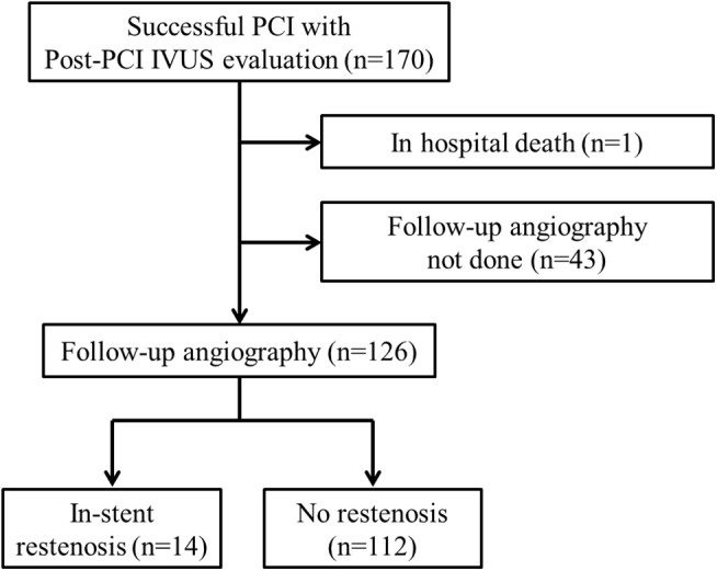
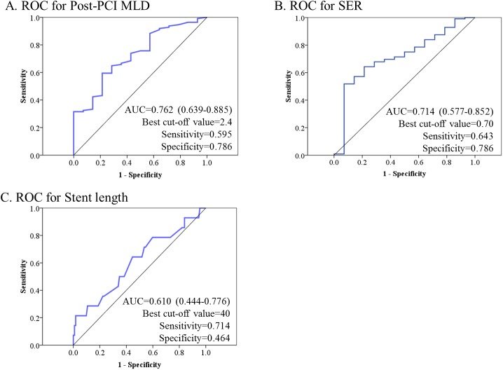
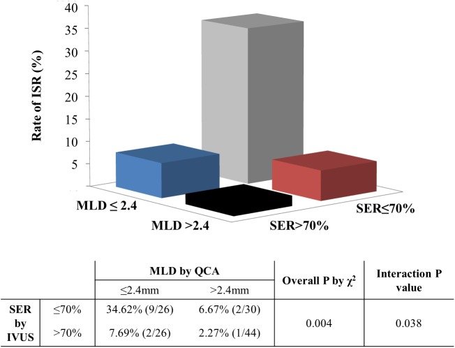
그림 출처: PubMed Central 공개 논문 (PMC4605613)
Conflict of interest statement
Competing Interests:The authors have declared that no competing interests exist.
그 밖의 정보
- 출판 형태
- Journal Article
- 저널
- PloS one · eISSN 1932-6203
- 전자 출판
- 2015-10-14
- MeSH
- Aged · Coronary Angiography* · Coronary Restenosis / diagnostic imaging / etiology · Drug-Eluting Stents / adverse effects · Echocardiography* · Endosonography* · Female · Humans · Male · Middle Aged · Percutaneous Coronary Intervention / adverse effects
- 참고문헌
- 29편