Department of Internal Medicine, Seoul National University Boramae Hospital, Seoul National University College of Medicine, Seoul, Republic of Korea.
Department of Internal Medicine and Liver Research Institute, Seoul National University College of Medicine, Seoul, Republic of Korea.
Department of Internal Medicine and Healthcare Research Institute, Healthcare System Gangnam Center, Seoul National University Hospital, Seoul, Republic of Korea.
Department of Internal Medicine and Liver Research Institute, Seoul National University College of Medicine, Seoul, Republic of Korea. jooskim@snu.ac.kr.
Department of Internal Medicine and Healthcare Research Institute, Healthcare System Gangnam Center, Seoul National University Hospital, Seoul, Republic of Korea. jooskim@snu.ac.kr.
Abstract
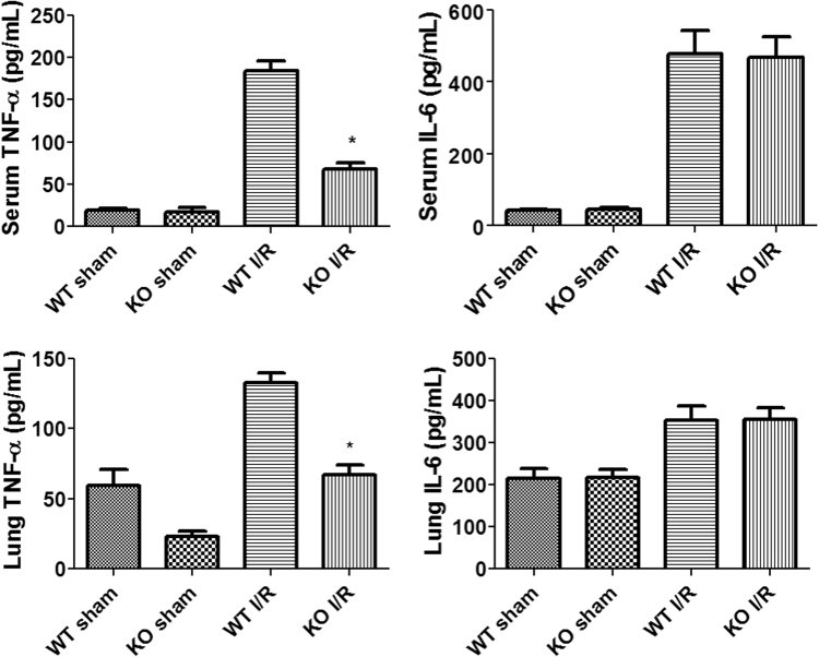
Intestinal ischemia-reperfusion (I/R) may cause acute systemic and lung inflammation. However, the detailed mechanism of this inflammatory cascade has not been fully elucidated. Inactive rhomboid protein 2 (iRhom2) is essential for the maturation of TNF-α converting enzyme (TACE), which is required for TNF-α secretion. We evaluated the role of iRhom2 in a mouse model of intestinal I/R using iRhom2 knockout (KO) and wild-type (WT) mice. Lung injury following intestinal I/R was significantly attenuated in iRhom2 KO mice compared with WT mice. After intestinal I/R, lungs from iRhom2 KO mice showed significantly lower myeloperoxidase (MPO) activity and markedly reduced cell apoptosis associated with a decreased level of active caspase 3 and decreased TUNEL staining compared with lungs from WT mice. TNF-α levels were elevated in the serum and lungs of WT mice with intestinal I/R and significantly reduced in iRhom2 KO mice with intestinal I/R. iRhom2 may play a critical role in the pathogenesis of acute lung injury (ALI) after intestinal I/R and thus may be a novel therapeutic target for ALI after intestinal I/R injury.
Figures
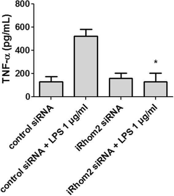
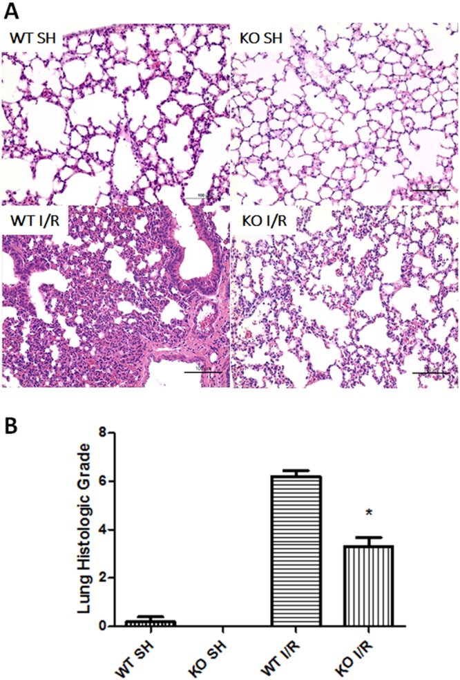
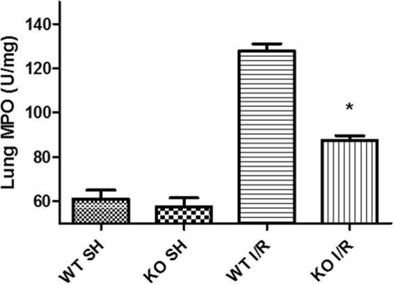
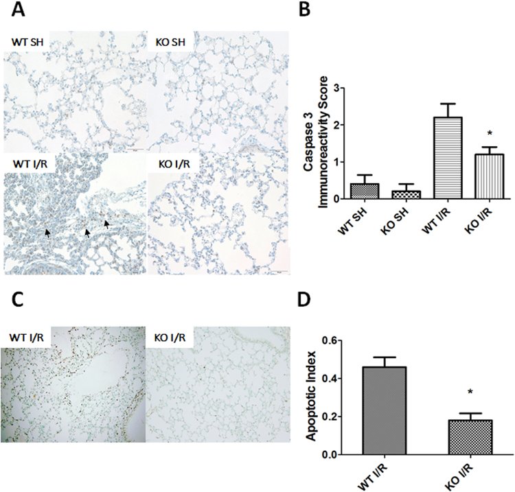
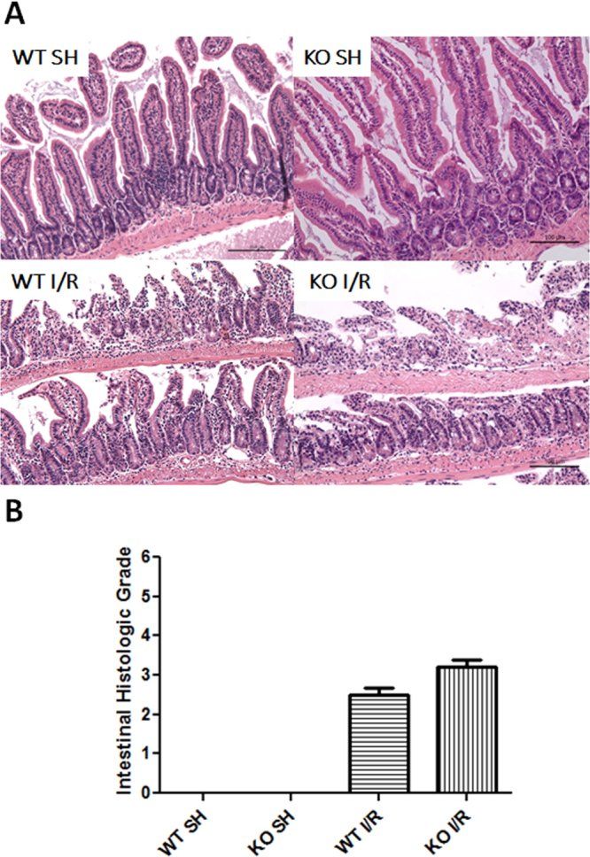
Figure 1siRNA-mediated iRhom2 knockdown inhibits TNF-α secretion in LPS-stimulated macrophages. RAW264.7 cells were transfected with iRhom2 or control siRNA before LPS stimulation, and TNF-α secretion by LPS-stimulated RAW264.7 cells was measured by ELISA. TNF-α secretion by LPS-stimulated macrophages was significantly inhibited by iRhom2 knockdown. *P < 0.05 versus the control siRNA + LPS group.Figure 2iRhom2 deficiency ameliorates lung injury after intestinal ischemia-reperfusion (I/R). (A) Representative histological sections of hematoxylin and eosin (H&E)-stained lung tissues from wild-type (WT) mice and iRhom2 knockout (KO) mice are shown (x200). Scale bars: 100 μm. The iRhom2 KO and WT mice were randomized into an intestinal I/R group (each n = 10) and a sham laparotomy group (each n = 5) as follows: KO SH (sham-operated KO group), WT SH (sham-operated WT group), KO I/R (KO with intestinal I/R group), and WT I/R (WT with intestinal I/R group). Lung injury following intestinal I/R was significantly attenuated in the iRhom2 KO mice compared with the WT mice. (B) Lung injury was assessed by using a modified scoring system with three criteria: alveolar edema/exudates, hemorrhage and interstitial/alveolar cellular infiltration. *P < 0.05 versus the WT I/R group.Figure 3iRhom2 deficiency alleviates lung neutrophil infiltration after intestinal ischemia-reperfusion (I/R). The myeloperoxidase (MPO) activity in the lung tissues was measured to quantitate the accumulation of neutrophils in the lung. The lung MPO activity was significantly decreased in the iRhom2 knockout (KO) mice compared with the wild-type (WT) mice after intestinal I/R. *P < 0.05 versus the WT I/R group.Figure 4iRhom2 deficiency reduces lung cell apoptosis after intestinal ischemia-reperfusion (I/R). (A) Representative images of active caspase 3 immunohistochemical staining in lung tissues (cleaved caspase 3 [diluted 1:1000]) are shown (x400). Scale bars: 50 μm. After intestinal I/R injury, the active caspase 3 protein levels were substantially reduced in the iRhom2 knockout (KO) mice compared with the wild-type (WT) mice. The arrows indicate active caspase 3-positive cells. (B) The immunoreactivity score of caspase 3 in the lung tissues was significantly decreased in the iRhom2 KO mice compared with the WT mice. The immunoreactivity of caspase 3 was assessed by determining the percentage of positive cells using a visual scoring system and was classified as 0 (no staining), 1+ (<10%), 2+ (10–30%), 3+ (31–60%) and 4+ (61–100%). *P < 0.05 versus the WT I/R group. (C) Terminal deoxynucleotidyl transferase dUTP nick end labeling (TUNEL) assays of the lungs after intestinal I/R in the WT and iRhom2 KO mice are shown (x200). TUNEL staining was reduced in the iRhom2 KO mice compared with the WT mice after intestinal I/R. The black-brown coloring indicates positive staining, and green indicates the contrast background staining. (D) The apoptotic index was also markedly reduced in the lungs of iRhom2 KO mice. The apoptotic index was calculated based on the ratio of TUNEL-reactive cells to the total number of cells in each of three randomly selected, nonadjacent fields. *P < 0.05 versus the WT I/R group.Figure 5iRhom2 deficiency does not affect intestinal injury after intestinal ischemia-reperfusion (I/R). (A) Representative histological sections of H&E-stained intestines from the wild-type (WT) and iRhom2 knockout (KO) mice are shown (x100). Scale bars: 100 μm. The intestinal injury was not different between the iRhom2 WT and KO mice. (B) The severity of the intestinal injury was assessed using a histological grading system based on the following criteria (score: 0–5): preservation of the villi, epithelial degeneration/necrosis, erosion or ulceration, and mucin depletion.Figure 6Alterations to inflammatory cytokines after intestinal ischemia-reperfusion (I/R). Inflammatory cytokine proteins (TNF-α and IL-6) in the serum and lung were measured by ELISA. *P < 0.05 versus the WT I/R group.