
Int J Infect Dis. 2020;97:313-321.
Aberrant hyperactivation of cytotoxic T-cell as a potential determinant of COVID-19 severity.
제1저자 강창경, 한기찬, 김민지 · 교신저자 김항래, 오명돈
소속 (6)
- Department of Internal Medicine, Seoul National University College of Medicine, Seoul 03080, Republic of Korea.
- Department of Biomedical Sciences, Seoul National University College of Medicine, Seoul 03080, Republic of Korea; Department of Anatomy & Cell Biology, Seoul National University College of Medicine, Seoul 03080, Republic of Korea; BK21 Plus Biomedical Science Project, Seoul National University College of Medicine, Seoul 03080, Republic of Korea.
- Wide River Institute of Immunology, Seoul National University, Hongcheon 25159, Republic of Korea.
- Department of Internal Medicine, Seoul National University College of Medicine, Seoul 03080, Republic of Korea; Department of Internal Medicine, Seoul National University Bundang Hospital, Seongnam 13620, Republic of Korea.
- Department of Biomedical Sciences, Seoul National University College of Medicine, Seoul 03080, Republic of Korea; Department of Anatomy & Cell Biology, Seoul National University College of Medicine, Seoul 03080, Republic of Korea; BK21 Plus Biomedical Science Project, Seoul National University College of Medicine, Seoul 03080, Republic of Korea; Wide River Institute of Immunology, Seoul National University, Hongcheon 25159, Republic of Korea; Medical Research Institute, Seoul National University College of Medicine, Seoul 03080, Republic of Korea. Electronic address: hangrae2@snu.ac.kr.
- Department of Internal Medicine, Seoul National University College of Medicine, Seoul 03080, Republic of Korea. Electronic address: mdohmd@snu.ac.kr.
Abstract
OBJECTIVES We hypothesized that immune response may contribute to progression of coronavirus disease-19 (COVID-19) at the second week of illness. Therefore, we compared cell-mediated immune (CMI) responses between severe and mild COVID-19 cases.
METHODS We examined peripheral blood mononuclear cells of laboratory-confirmed COVID-19 patients from their first and third weeks of illness. Severe pneumonia was defined as an oxygen saturation ≤93% at room air. Expressions of molecules related to T-cell activation and functions were analyzed by flow cytometry.
RESULTS The population dynamics of T cells at the first week were not different between the two groups. However, total numbers of CD4+ and CD8+ T cells tended to be lower in the severe group at the third week of illness. Expressions of Ki-67, PD-1, perforin, and granzyme B in CD4+ or CD8+ T cells were significantly higher in the severe group than in the mild group at the third week. In contrast to the mild group, the levels of their expression did not decrease in the severe group.
CONCLUSIONS Severe COVID-19 had a higher degree of proliferation, activation, and cytotoxicity of T-cells at the late phase of illness without cytotoxic T-cell contraction, which might contribute to the development of severe COVID-19.
Copyright © 2020 The Author(s). Published by Elsevier Ltd.. All rights reserved.
Keywords
COVID-19ContractionCytotoxic T cellGranzyme BPerforinSeverity
Figures
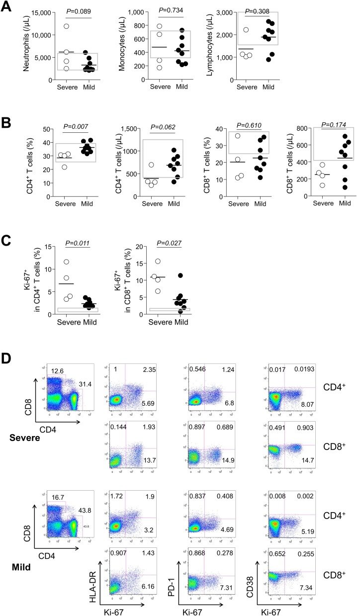
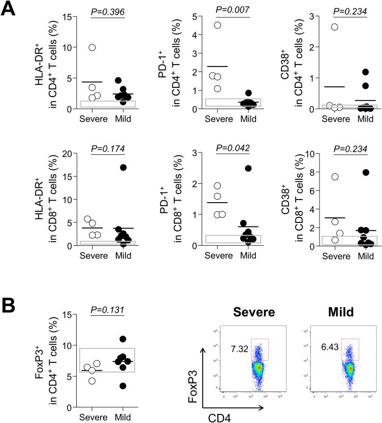
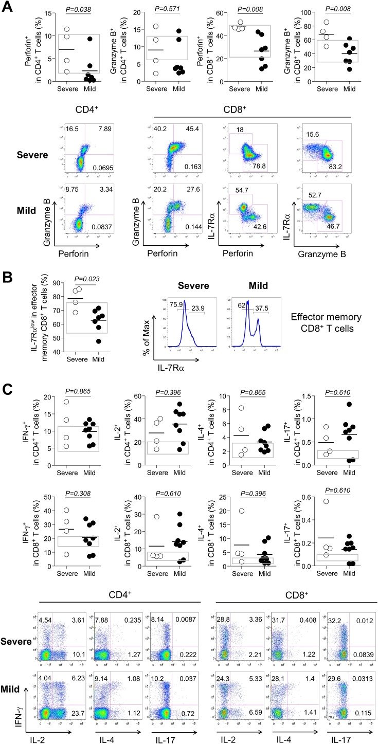
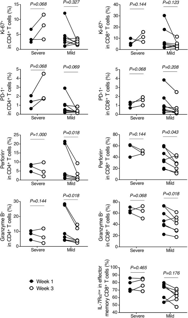
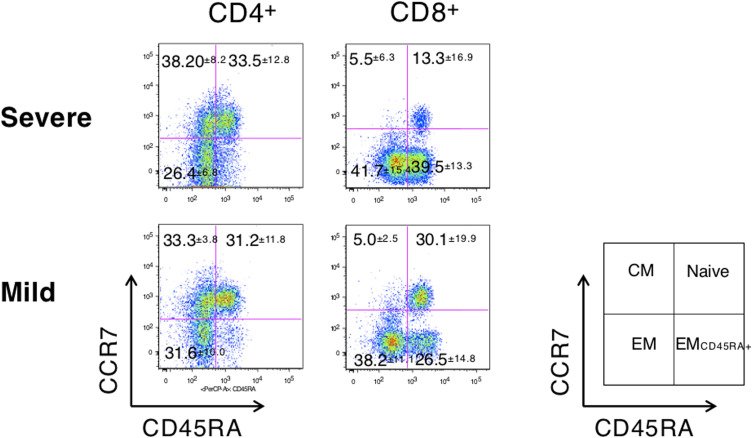
그림 출처: PubMed Central 공개 논문 (PMC7261468)
그 밖의 정보
- 출판 형태
- Journal Article
- 저널
- International journal of infectious diseases : IJID : official publication of the International Society for Infectious Diseases · eISSN 1878-3511
- 전자 출판
- 2020-05-31
- MeSH
- Adult · Aged · Aged, 80 and over · Betacoronavirus · CD4-Positive T-Lymphocytes / immunology · CD8-Positive T-Lymphocytes / immunology · COVID-19 · Coronavirus Infections / immunology / physiopathology · Cytokines / immunology · Female · Granzymes · Humans · Immunity, Cellular* · Ki-67 Antigen · Leukocytes, Mononuclear / immunology · Lymphocyte Activation* · Lymphocyte Count · Male · Middle Aged · Pandemics · Perforin · Pneumonia, Viral / immunology / physiopathology · Programmed Cell Death 1 Receptor · SARS-CoV-2 · T-Lymphocytes, Cytotoxic / immunology
- Substances
- Cytokines · Ki-67 Antigen · MKI67 protein, human · PDCD1 protein, human · Programmed Cell Death 1 Receptor · Perforin · GZMB protein, human · Granzymes
- 참고문헌
- 29편