From the Cardiovascular Center and Department of Internal Medicine, Seoul National University Hospital, Republic of Korea (I.-C.H., J.-H.K., J.P., H.-M.Y., H.-J.C., H.-S.K.).
National Leading Laboratory for Cardiovascular Stem Cell, Seoul National University College of Medicine, Republic of Korea (I.-C.H., J.-Y.K., J.-H.K., J.-E.L., J.-Y.S., J.-W.L., J.P., H.-M.Y., H.-J.C., H.-S.K.).
Strategic Center of Cell and Bio Therapy for Heart, Diabetes, and Cancer, Seoul National University Hospital, Republic of Korea (I.-C.H., J.-Y.K., J.-E.L., J.-W.L., H.-M.Y., H.-J.C., H.-S.K.).
R&D Campus, LG Chem/Ltd., Daejeon, Republic of Korea (S.-H.K.).
From the Cardiovascular Center and Department of Internal Medicine, Seoul National University Hospital, Republic of Korea (I.-C.H., J.-H.K., J.P., H.-M.Y., H.-J.C., H.-S.K.) hyosoo@snu.ac.kr.
and Department of Molecular Medicine and Biopharmaceutical Science, Seoul National University, Republic of Korea (H.-S.K.).
Abstract
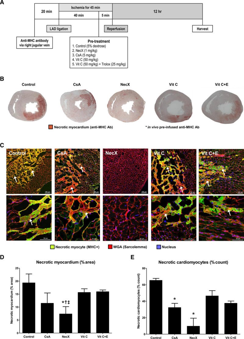
Opening of mitochondrial permeability transition pore and Ca2+ overload are main contributors to myocardial ischemia-reperfusion injury, which paradoxically causes a wide variety of myocardial damage. We investigated the protective role of a novel necrosis inhibitor (NecroX-7; NecX) against myocardial ischemia-reperfusion injury using in vitro and in vivo models. H9C2 rat cardiomyoblasts and neonatal cardiomyocytes were exposed to hypoxia-reoxygenation stress after pre-treatment with NecX, vitamin C, a combination of vitamin C and E, N-acetylcysteine, an apoptosis inhibitor (Z-VAD-fmk), or cyclosporine A. The main mechanism of cell death after hypoxia-reoxygenation stress was not apoptosis but necrosis, which was prevented by NecX. Protective effect of NecX was based on its potent reactive oxygen species scavenging activity, especially on mitochondrial reactive oxygen species. NecX preserved mitochondrial membrane potential through prevention of Ca2+ influx and inhibition of mitochondrial permeability transition pore opening, which was more potent than that by cyclosporine A. Using Sprague-Dawley rats exposed to myocardial ischemia for 45 minutes followed by reperfusion, we compared therapeutic efficacies of NecX with cyclosporine A, vitamin C, a combination of vitamin C and E, and 5% dextrose, each administered 5 minutes before reperfusion. NecX markedly inhibited myocardial necrosis and reduced fibrotic area to a greater extent than did cyclosporine A and other treated groups. In addition, NecX preserved systolic function and prevented pathological dilatory remodeling of left ventricle. The novel necrosis inhibitor has a significant protective effect against myocardial ischemia-reperfusion injury through inhibition of mitochondrial permeability transition pore opening, indicating that it is a promising candidate for cardioprotective adjunctive measure on top of reperfusion therapy.
Figure 1.Protection of cardiomyocytes from necrosis after hypoxia–reoxygenation stress by NecX. A, Chemical structure of the novel necrosis inhibitor, 7-amino-indole (NecroX-7; NecX). B, Experimental protocol. C, Representative figures of H9C2 rat cardiomyoblasts stained with fluorescein diacetate (FDA)/propidium iodide (PI). D, Percent of dead cells by FDA/PI staining. E, Contour plots of annexin-V vs PI, showing necrotic cells in red color (annexin-V/PI −/+; upper left) and apoptotic cells in blue color (annexin-V/PI +/+; upper right) among H9C2 rat cardiomyoblasts. F, Percentages of viable, apoptotic, and necrotic cells calculated. *P<0.05 vs vehicle, and †P<0.05 vs NecX. Bars represent the mean±SEM from 3 independent experiments. NAC indicates N-acetylcysteine; NecX, necrosis inhibitor; Veh, vehicle; Vit C, vitamin C; and Vit E, vitamin E.Figure 2.Activation of necrosis signaling pathways after hypoxia–reoxygenation stress in H9C2 rat cardiomyoblasts. A, Two cleaved forms of PARP1 (poly[ADP-ribose] polymerase 1); apoptotic fragment of cleaved PARP1 (89 kDa) and necrotic fragment of cleaved PARP1 (55 kDa). Activation of apoptosis pathways was assessed by (B) Western blot of cleaved caspase-3 and (C) the luminescence assay for caspase-3 activity. D, Western blot analyses of key molecules involved in necrosis; RIP1 (receptor-interacting protein 1), RIP3, phospho-p38 MAPK (mitogen-activated protein kinase), total p38 MAPK, phospho-JNK (Jun N-terminal kinase), and total JNK. *P<0.05 vs vehicle, and †P<0.05 vs SP (staurosporine; apoptosis inducer). Bar represents the mean±SEM from 4 independent experiments. Con indicates control; SP, staurosporine; and SS, serum starvation.Figure 3.Mitochondrial effect of the necrosis inhibitor. A and B, Mitochondria-specific reactive oxygen species (orange fluorescence) was measured in H9C2 rat cardiomyoblasts under hypoxia–reoxygenation stress using dihydrorhodamine 123 assay. C, Mitochondrial membrane potential (Δψm) was measured using tetramethylrhodamine ethyl ester (TMRE; orange fluorescence) probe. D, Flow cytometry histograms of TMRE fluorescence. E, Mitochondrial membrane potential (Δψm) was measured in neonatal rat cardiomyocytes. The cells pre-treated with NecX showed significantly preserved high Δψm (orange fluorescence), whereas vehicle, vitamin C, or apoptosis inhibitor demonstrated low Δψm. F, Flow cytometry dot plots of JC-1 monomers (low Δψm; right lower) vs J-aggregates (high Δψm; right upper). The NecX-treated cells demonstrated higher mitochondrial membrane potentials while vehicle, vitamin C, and Z-VAD-fmk did not maintain normal mitochondrial membranes. *P<0.05 vs vehicle, and †P<0.05 vs NecX. Bars represent the mean±SEM from 3 independent experiments.Figure 4.Inhibition of mitochondrial permeability transition pore (mPTP) opening and prevention of calcium influx into mitochondria. A, The opening of mPTP was directly assessed by using calcein-AM (green fluorescence) with addition of cobalt chloride for mitochondrial localization of calcein-AM fluorescence. The cells were costained with MitoTracker (red fluorescence) and Hoechst 33342 (blue fluorescence) to demonstrate the mitochondria and nucleus, respectively. B, Time courses of changes in calcein-AM fluorescence intensity. Bars represent the mean±SEM. C, Two vertical columns depict the changes of mitochondrial Ca2+ level (Rhod-2 fluorescence; red) and mitochondrial shapes (MitoTracker; green) according to time course. CsA indicates cyclosporine A.Figure 5.Myocardial necrosis after ischemia–reperfusion injury. A, Experimental protocol. B, Heart tissue sections showing necrotic zones stained in dark-brown color by the use of anti–myosin heavy chain (MHC) antibody. C, Results from immunohistochemistry of heart tissue sections showing necrotic cardiomyocytes in green fluorescence and intact cardiomyocytes in red fluorescence (staining the sarcolemma) and blue fluorescence (staining nucleus). D, Necrotic areas expressed as the percent of total left ventricular areas. E, Necrotic cardiomyocytes expressed as the percent of total myocytes. *P<0.05 vs control, †P<0.05 vs Vit C, and ‡P<0.05 vs Vit C+E; n=5 per group. Bars represent the mean±SEM. CsA indicates cyclosporine A; and Vit, vitamin.Figure 6.Myocardial fibrosis and echocardiographic parameters after ischemia–reperfusion (I/R) injury. A, Experimental protocol. B, Representative Masson trichrome–stained slides of the myocardium sections from each treated group obtained at 14 d after myocardial I/R injury. C and D, Comparison of the areas of fibrosis and the percentages of fibrotic myocardium to the total myocardium. *P<0.05 vs control, and †P<0.05 vs cyclosporine A (CsA); n=5 per group. E and F, Echocardiographic parameters including left ventricular ejection fraction and end-diastolic dimension. *P<0.05 vs control, and †P<0.05 vs CsA; n=10 per group. Bars represent the mean±SEM. LAD indicates left anterior descending coronary artery.