
Medicine (Baltimore). 2017;96(5):e5926.
Serum interleukin-6 is associated with pancreatic ductal adenocarcinoma progression pattern.
제1저자 김형우 · 교신저자 황진혁
소속 (1)
- Department of Internal Medicine, Seoul National University College of Medicine, Seoul National University Bundang Hospital, Seongnam, Korea.
Abstract
Several reports showed that interleukin-6 (IL-6) or -8 (IL-8) might be useful inflammatory biomarkers for pancreatic ductal adenocarcinoma (PDAC), although these clinical impact is still open to debate. The aim of this study was to elucidate whether serum levels of IL-6 and IL-8 at diagnosis could predict the tumor progression pattern of PDAC, especially in extensive hepatic metastasis.According to the tumor burden of hepatic metastasis at the last follow-up, tumor progression pattern was defined as follows: no or limited (unilobar involvement and 5 or less in the within liver, limited group) and extensive hepatic metastasis (bilobar or more than 5, progressed group). Fifty-three PDAC patients with initially no or limited hepatic metastasis were enrolled retrospectively.Around 42 (79.2%) were included in the limited and 11 (20.8%) in the progressed group. The median serum level of IL-6 in the progressed group was elevated significantly compared with the limited group. However, the median serum level of IL-8 was not. Furthermore, multivariate analysis revealed that the elevated serum level of IL-6 was an independent risk factor for progression to extensive hepatic metastasis (odds ratio 1.928, 95% confidence interval 1.131-3.365, P = 0.019), but IL-8 was not. However, higher IL-6 did not predict shorter survival.High serum IL-6 can be an independent risk factor for progression to extensive hepatic metastasis in PDAC patients.
Figures
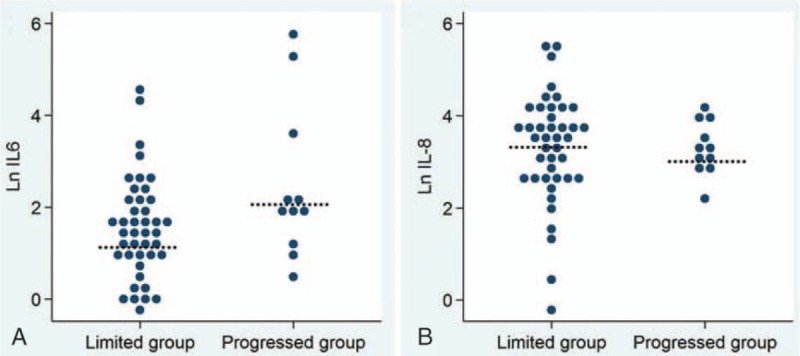
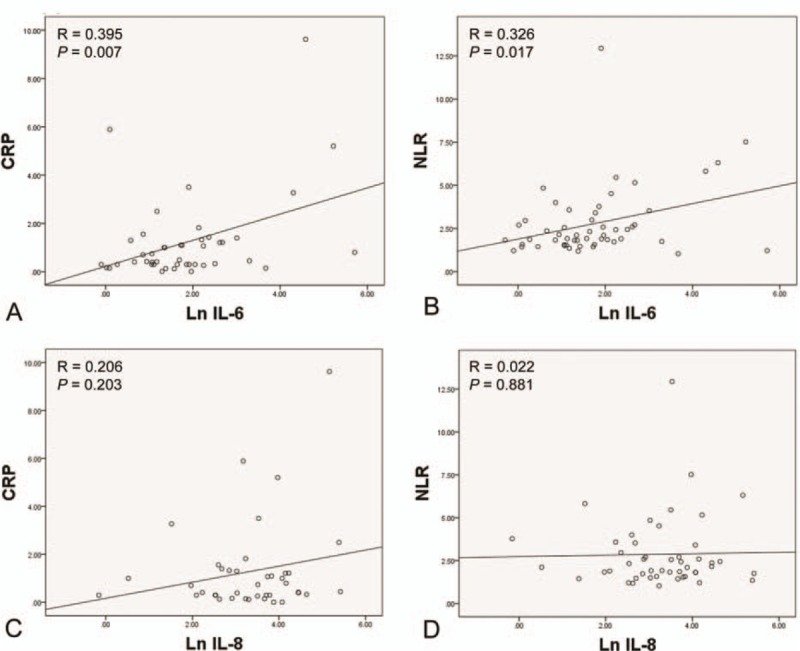
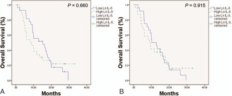
그림 출처: PubMed Central 공개 논문 (PMC5293435)
Conflict of interest statement
The authors have no conflicts of interest to disclose.
그 밖의 정보
- 출판 형태
- Journal Article, Observational Study
- 저널
- Medicine · eISSN 1536-5964
- MeSH
- Aged · Biomarkers, Tumor · Carcinoma, Pancreatic Ductal / blood / mortality / pathology · Disease Progression · Female · Humans · Interleukin-6 / blood · Interleukin-8 / blood · Liver Neoplasms / secondary · Male · Middle Aged · Pancreatic Neoplasms / blood / mortality / pathology · Prognosis · Retrospective Studies · Risk Factors
- Substances
- Biomarkers, Tumor · Interleukin-6 · Interleukin-8
- 참고문헌
- 24편