
Gut Liver. 2013;7(3):371-6.
A Case of Common Bile Duct Cancer That Completely Responded to Combination Chemotherapy of Gemcitabine and TS-1.
제1저자 임주현 · 교신저자 류지곤
소속 (1)
- Department of Internal Medicine and Liver Research Institute, Seoul National University College of Medicine, Seoul, Korea.
Abstract
Common bile duct (CBD) cancer is a relatively rare malignancy that arises from the biliary epithelium and is associated with a poor prognosis. Here, we report a case of advanced metastatic CBD cancer successfully treated by chemotherapy with gemcitabine combined with S-1 (tegafur+gimeracil+oteracil). A 65-year-old male presented with pyogenic liver abscess. After antibiotic therapy and percutaneous drainage, follow-up computed tomography (CT) showed an enhanced nodule in the CBD. Biopsy was performed at the CBD via endoscopic retrograde cholangiopancreatography, which showed adenocarcinoma. Additional CT and magnetic resonance imaging showed multiple small nodules in the right hepatic lobe, which were confirmed as metastatic adenocarcinoma by sono-guided liver biopsy. The patient underwent combination chemotherapy with gemcitabine and S-1. After nine courses of chemotherapy, the hepatic lesion disappeared radiologically. Pylorus-preserving pancreaticoduodenectomy was performed, and no residual tumor was found in the resected specimen. Three weeks after the operation, the patient was discharged with no complications. Through 3 months of follow-up, no sign of recurrence was observed on CT scan. Gemcitabine combined with S-1 may be a highly effective treatment for advanced cholangiocarcinoma.
Keywords
CholangiocarcinomaGemcitabineS-1
Figures

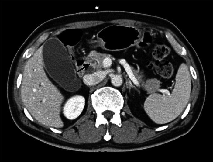

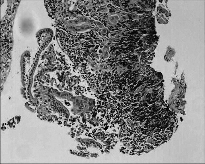
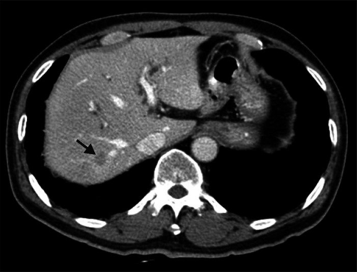
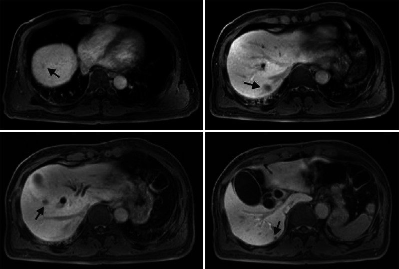
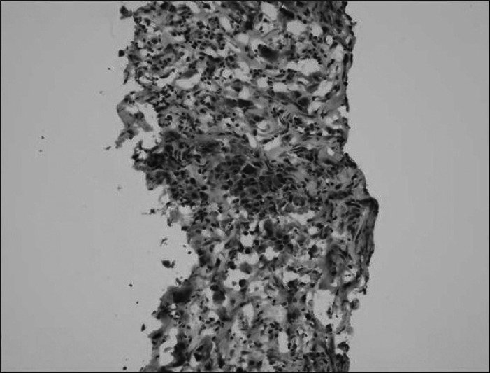

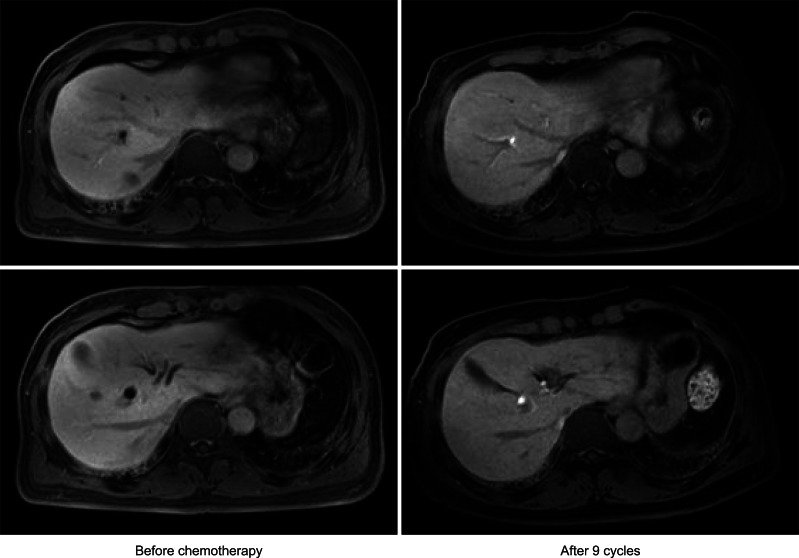
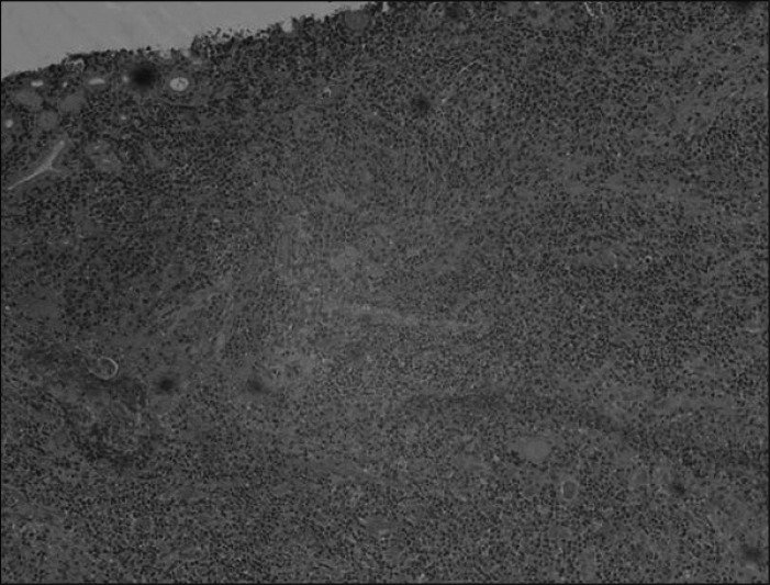

그림 출처: PubMed Central 공개 논문 (PMC3661972)
Conflict of interest statement
No potential conflict of interest relevant to this article was reported.
그 밖의 정보
- 출판 형태
- Journal Article
- 저널
- Gut and liver · ISSN 1976-2283
- 전자 출판
- 2013-05-13
- 참고문헌
- 20편