
Eur Heart J Digit Health. 2026;7(3):ztag028.
Deep learning-based multi-view echocardiographic framework for comprehensive diagnosis of pericardial disease.
제1저자 정시현, 문인태 · 교신저자 윤연이
소속 (6)
- Department of Internal Medicine, Graduate School of Medical Science, Brain Korea 21 Project, Yonsei University College of Medicine, Seoul, Republic of Korea.
- Division of Cardiology, Department of Internal Medicine, Uijeongbu Eulji Medical Center, Eulji University College of Medicine, Uijeongbu, Republic of Korea.
- Ontact Health Inc., Seoul, Republic of Korea.
- Division of Cardiology, Severance Cardiovascular Hospital, Yonsei University College of Medicine, Yonsei University Health System, Seoul, Republic of Korea.
- Cardiovascular Center and Division of Cardiology, Department of Internal Medicine, Seoul National University Bundang Hospital, Seongnam, Gyeonggi, Republic of Korea.
- Department of Internal Medicine, Seoul National University College of Medicine, Seoul, Republic of Korea.
Abstract
AIMS Pericardial disease spans a wide spectrum from small effusions to life-threatening tamponade or constriction. Transthoracic echocardiography (TTE) is the main diagnostic tool, but its interpretation is limited by operator dependence and incomplete functional assessment. Existing deep learning (DL) models focus mainly on effusion detection, lacking broader evaluation.
METHODS AND RESULTS We developed a DL-based framework that performs sequential assessment of pericardial disease: (i) morphological features, including effusion amount (normal/small/moderate/large) and pericardial thickening/adhesion (yes/no), from five B-mode views, and (ii) haemodynamic significance (yes/no), incorporating Doppler and inferior vena cava measurements. The developmental dataset comprises 2253 TTEs from multiple Korean institutions (225 for internal testing), and the independent external test set consists of 274 TTEs. In the internal test set, diagnostic accuracy was 81.8-97.3% for effusion, 91.6% for thickening/adhesion, and 86.2% for haemodynamic significance. External test set accuracy was 80.3-94.2%, 94.5%, and 85.5%, respectively. Area under the receiver operating curves for the three tasks were 0.92-0.99, 0.90, and 0.79 internally, and 0.95-0.98, 0.85, and 0.76 externally. Sensitivity for thickening/adhesion and haemodynamic significance improved from 66.7% to 77.3%, and 68.8% to 80.8%, respectively, when poor image quality were excluded. Similar performance gains were observed in subgroups with complete target views and a higher number of available video clips.
CONCLUSION This study presents the first DL-based TTE model for broader pericardial disease evaluation, integrating morphological with supportive functional assessments. The proposed framework demonstrated strong generalizability and aligned with the real-world diagnostic workflow. However, caution is warranted when interpreting results under suboptimal imaging conditions.
© The Author(s) 2026. Published by Oxford University Press on behalf of the European Society of Cardiology.
Keywords
Artificial IntelligenceDeep learningEchocardiographyHaemodynamic assessmentPericardial disease
Figures
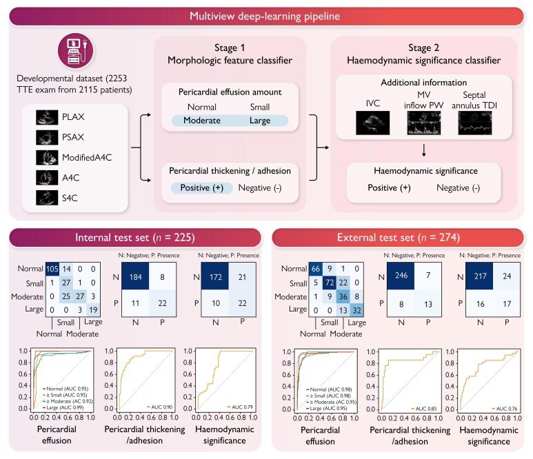
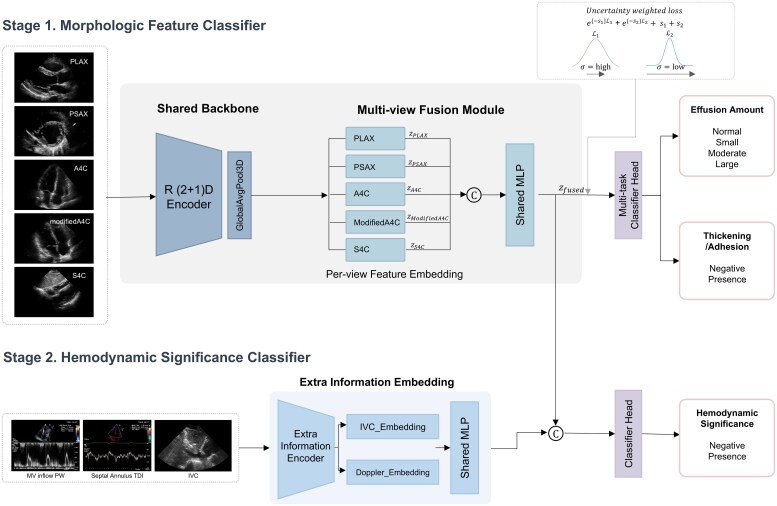
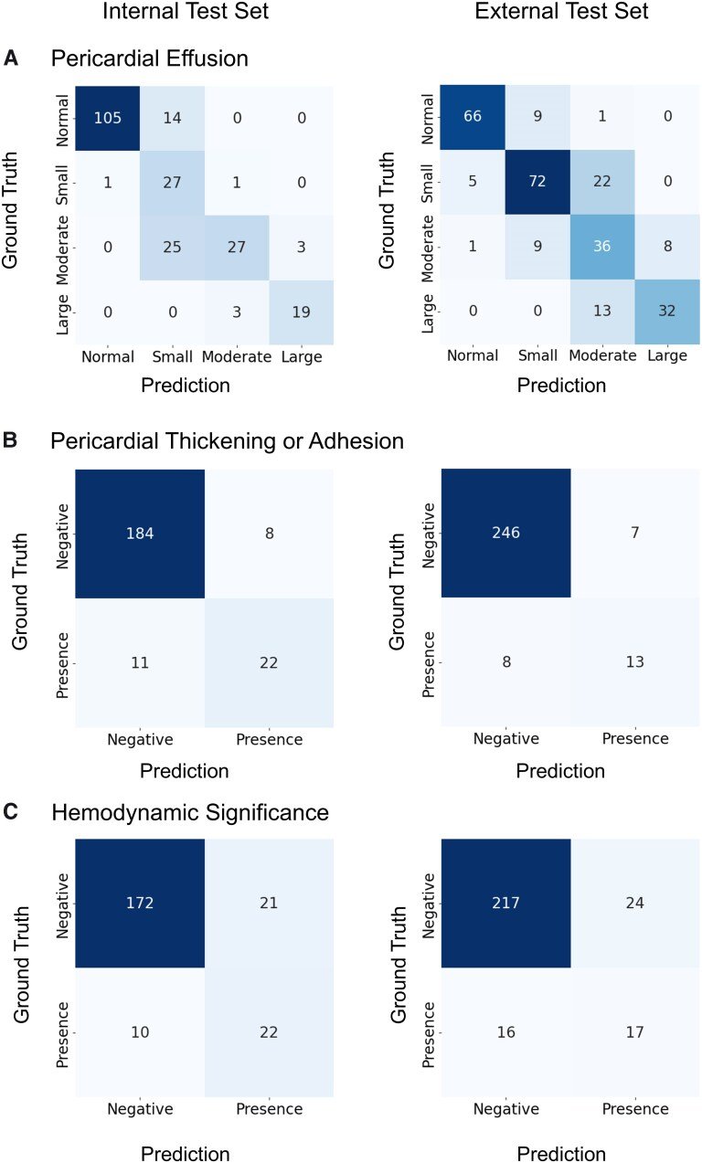
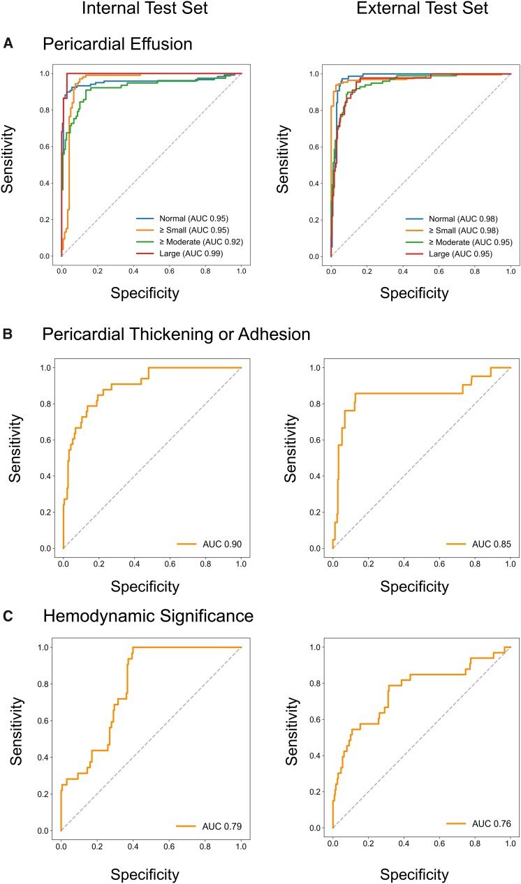
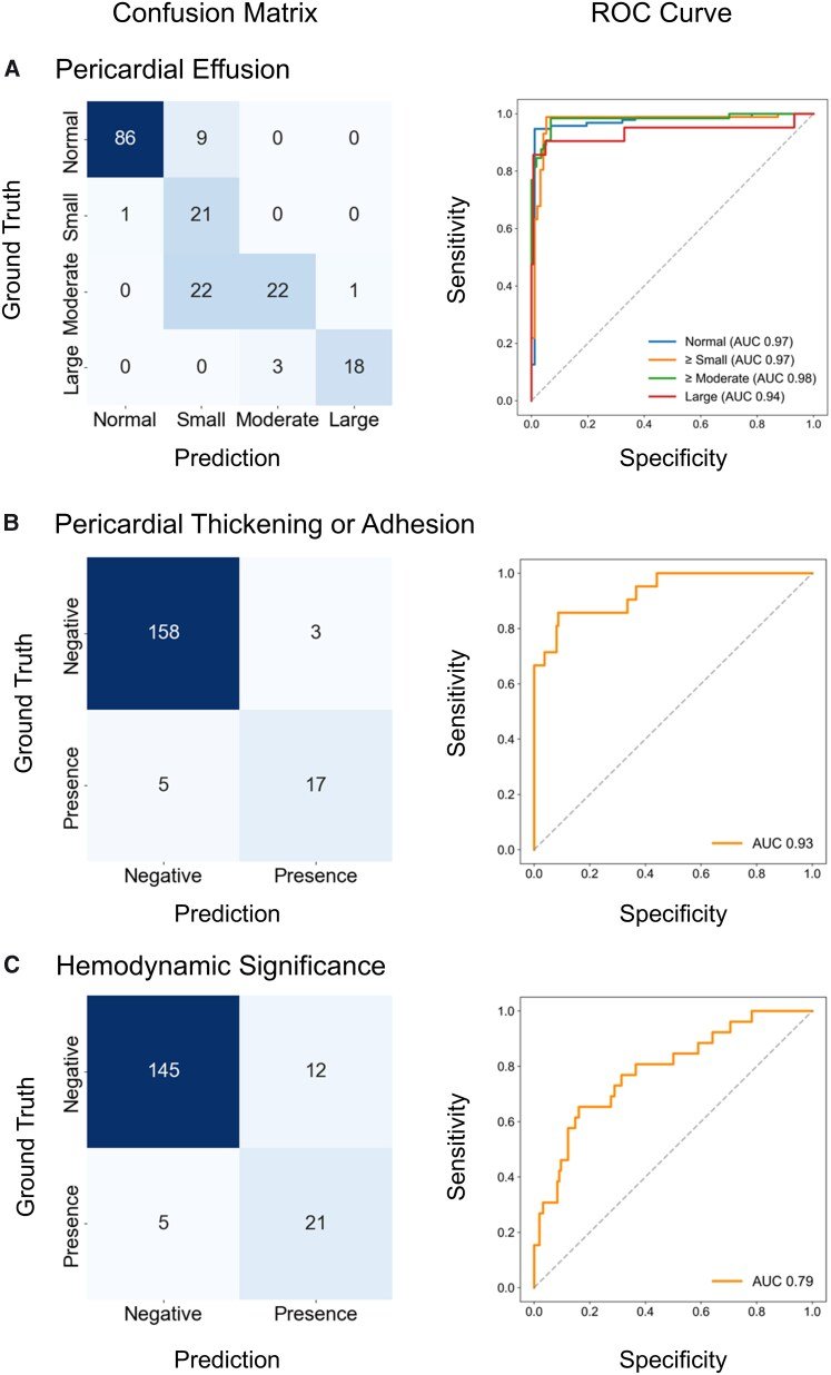
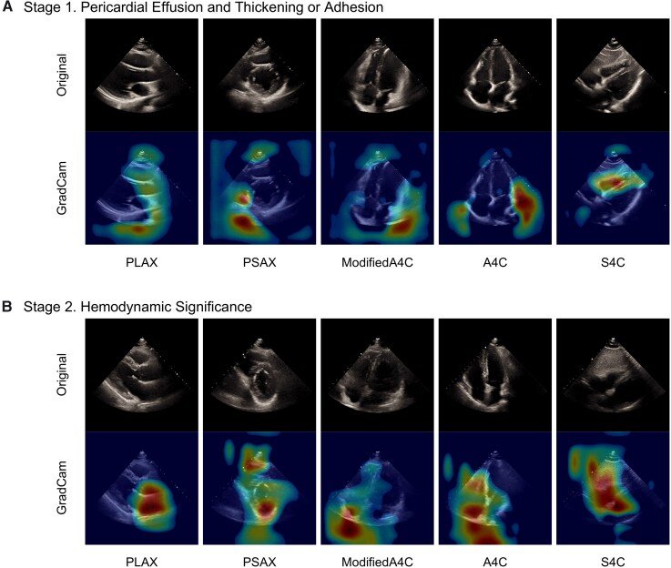
그림 출처: PubMed Central 공개 논문 (PMC12966503)
Conflict of interest statement
Conflict of interest: J.J., J.K., S.A.L., Y.J., and Y.E.Y. are currently affiliated with Ontact Health, Inc. Y.E.Y., and H.J.C. hold stock in Ontact Health, Inc. The other authors have no conflicts of interest to declare.
그 밖의 정보
- 출판 형태
- Journal Article
- 저널
- European heart journal. Digital health · eISSN 2634-3916
- 전자 출판
- 2026-03-07
- 참고문헌
- 23편