
Gut Liver. 2013;7(2):239-45.
Endoscopic papillectomy for benign ampullary neoplasms: how can treatment outcome be predicted?
제1저자 안동원 · 교신저자 류지곤
소속 (1)
- Department of Internal Medicine and Liver Research Institute, Seoul National University College of Medicine, Seoul, Korea.
Abstract
BACKGROUND/AIMS Endoscopic papillectomy is increasingly performed with curative intent for benign papillary tumors. This study was performed to identify factors that predict the presence of malignancy and affect endoscopic success.
METHODS We retrospectively analyzed the medical records of patients who received an endoscopic papillectomy for papillary adenoma from 2006 to 2009.
RESULTS A total of 43 patients received endoscopic papillectomy. The pathologic results after papillectomy revealed adenocarcinoma in five patients (12%), and the risk of malignancy was high in cases of large lesions, preprocedural pathology of high-grade dysplasia or high serum alkaline phosphatase. Endoscopic success was observed in 37 patients (86%) at the end of follow-up (mean duration, 10.4±9.6 months). The factor significantly affecting success was a complete resection at the initial papillectomy (p=0.007). Two patients experienced recurrence 10 and 32 months after the complete resection, but both achieved endoscopic success with repeated endoscopic treatment. Six patients with endoscopic failure received surgical resection.
CONCLUSIONS Endoscopic papillectomy is a safe and effective method for the curative resection of benign papillary tumors, especially when complete resection is achieved at the initial papillectomy. Follow-up with surveillance should be performed for at least 3 years because of the possible recurrence of tumors during these periods.
Keywords
AdenocarcinomaBenign papillary tumorEndoscopic sphincterotomyEndoscopic success
Figures

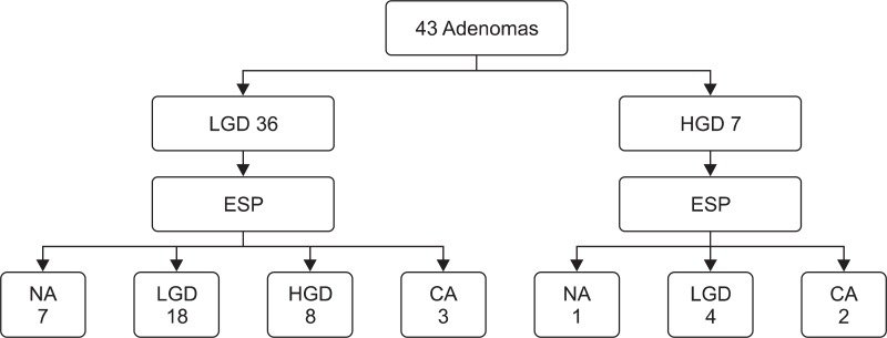
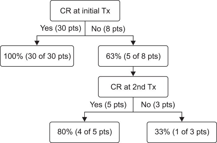
그림 출처: PubMed Central 공개 논문 (PMC3607780)
Conflict of interest statement
No potential conflict of interest relevant to this article was reported.
그 밖의 정보
- 출판 형태
- Journal Article
- 저널
- Gut and liver · ISSN 1976-2283
- 전자 출판
- 2013-03-14
- 참고문헌
- 26편