
Oncotarget. 2016;7(13):15901-14.
PD-L1 expression is associated with epithelial-mesenchymal transition in head and neck squamous cell carcinoma.
제1저자 옥찬영, 김세희 · 교신저자 김범석
소속 (6)
- Department of Internal Medicine, Seoul National University Hospital, Seoul, Korea.
- Department of Pathology, Seoul National University Hospital Seoul, Korea.
- Cancer Research Institute, Seoul National University College of Medicine, Seoul, Korea.
- Department of Radiation Oncology, Seoul National University Hospital, Seoul, South Korea.
- Department of Systems Biology, The University of Texas MD Anderson Cancer Center, Houston, Texas, USA.
- Department of Otorhinolaryngology, Seoul National University Hospital, Seoul, South Korea.
Abstract
Virus-associated malignancies and sarcomatoid cancers correlate with high PD-L1 expression, however, underlying mechanisms remain controversial. We evaluated the correlation between PD-L1 expression and epithelial-mesenchymal transition (EMT) in head and neck squamous cell carcinomas (HNSCC).Tumor tissues from 50 patients with HNSCC were evaluated for PD-L1 by immunohistochemistry, which showed 32 (64.0%) were PD-L1 positive (PD-L1+). Interestingly, PD-L1 expression was significantly associated with EMT (P = 0.010), as assessed by low E-cadherin and high vimentin expression. The overall survival of PD-L1+ patients with EMT features was significantly worse than those without EMT features (P = 0.007). In an independent validation cohort (N = 91), as well as in HNSCC cases of The Cancer Genome Atlas (TCGA) and the Cancer Cell Line Encyclopedia, high PD-L1 expression was also associated with the high probability of an EMT signature, referred from the GEO dataset, GSE4824. Survival analysis confirmed PD-L1+/EMT+ patients had a poorer prognosis than PD-L1+/EMT- patients in the TCGA cohort. PD-L1 positivity can thus be divided into two categories according to the absence or presence of EMT. PD-L1 expression is also independently associated with EMT features in HNSCC.
Keywords
PD-L1epithelial-mesenchymal transitionhead and neckp16squamous
Figures

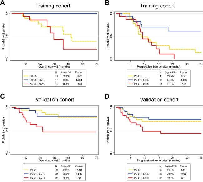
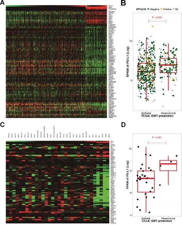
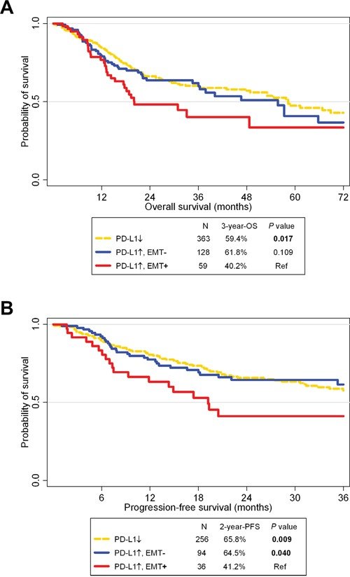
그림 출처: PubMed Central 공개 논문 (PMC4941285)
Conflict of interest statement
CONFLICTS OF INTEREST. All authors have no conflicts of interest to declare.
그 밖의 정보
- 출판 형태
- Journal Article
- 저널
- Oncotarget · eISSN 1949-2553
- MeSH
- Adolescent · Adult · Aged · B7-H1 Antigen / biosynthesis · Carcinoma, Squamous Cell / metabolism / mortality / pathology · Epithelial-Mesenchymal Transition / physiology · Female · Head and Neck Neoplasms / metabolism / mortality / pathology · Humans · Kaplan-Meier Estimate · Male · Middle Aged · Squamous Cell Carcinoma of Head and Neck · Young Adult
- Substances
- B7-H1 Antigen · CD274 protein, human
- 참고문헌
- 51편