Department of Internal Medicine, Maryknoll Hospital, Busan, Korea.
Abstract
Gentamicin nephrotoxicity is one of the most common causes of acute kidney injury (AKI). Hypoxia-inducible factor (HIF) is effective in protecting the kidney from ischemic and toxic injury. Increased expression of HIF-1α mRNA has been reported in rats with gentamicin-induced renal injury. We hypothesizd that we could study the role of HIF in gentamicin-induced AKI by modulating HIF activity. In this study, we investigated whether HIF activation had protective effects on gentamicin-induced renal tubule cell injury. Gentamicin-induced AKI was established in male Sprague-Dawley rats. Cobalt was continuously infused into the rats to activate HIF. HK-2 cells were pre-treated with cobalt or dimethyloxalylglycine (DMOG) to activate HIF and were then exposed to gentamicin. Cobalt or DMOG significantly increased HIF-1α expression in rat kidneys and HK-2 cells. In HK-2 cells, HIF inhibited gentamicin-induced reactive oxygen species (ROS) formation. HIF also protected these cells from apoptosis by reducing caspase-3 activity and the amount of cleaved caspase-3, and -9 proteins. Increased expression of HIF-1α reduced the number of gentamicin-induced apoptotic cells in rat kidneys and HK-2 cells. HIF activation improved the creatinine clearance and proteinuria in gentamicin-induced AKI. HIF activation also ameliorated the extent of histologic injury and reduced macrophage infiltration into the tubulointerstitium. In gentamicin-induced AKI, the activation of HIF by cobalt or DMOG attenuated renal dysfunction, proteinuria, and structural damage through a reduction of oxidative stress, inflammation, and apoptosis in renal tubular epithelial cells.
Figures
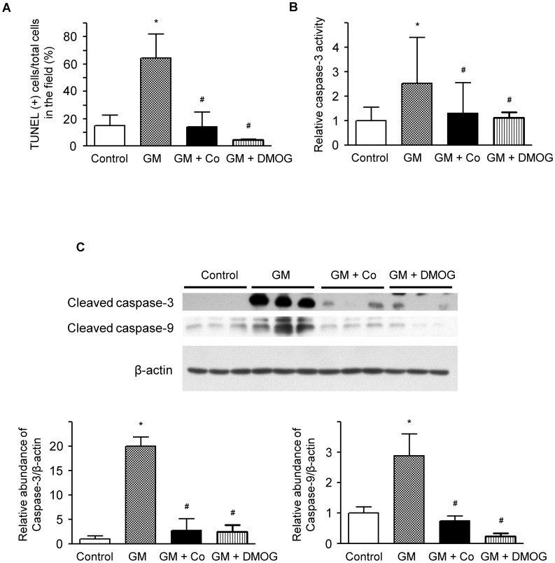
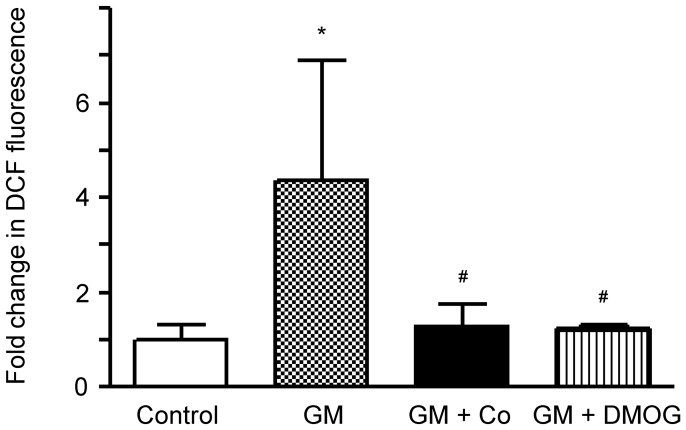
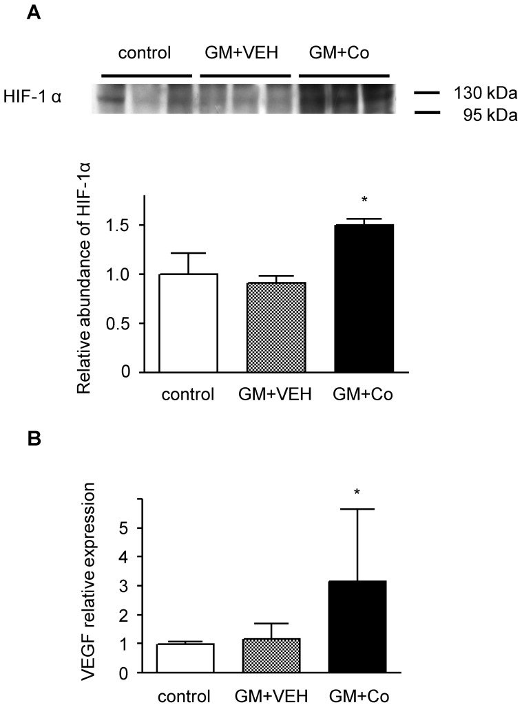
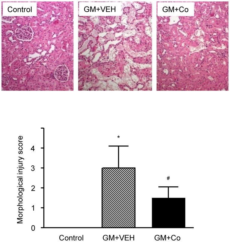
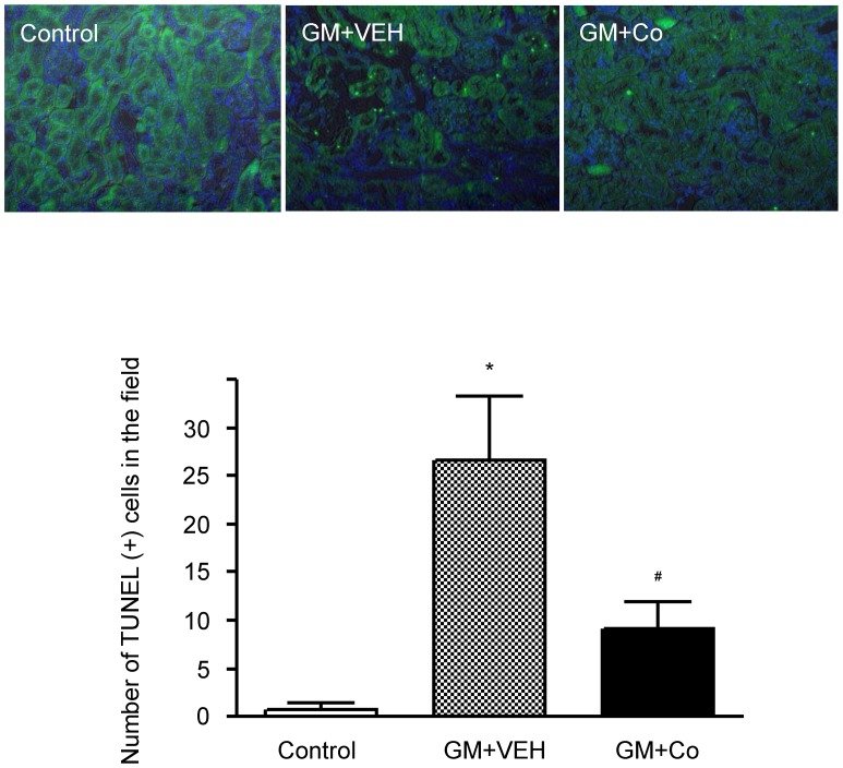
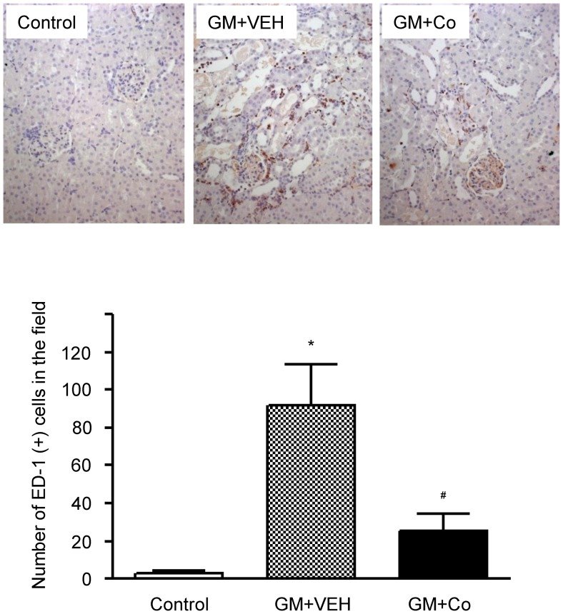
Figure 1 Activation of hypoxia-inducible factor (HIF) in HK-2 cells. Western blotting of HIF-1α in HK-2 cells. HK-2 cells were pre-treated with 150 µM of cobalt or 1 mM of DMOG for 24 h and then treated with 3 mM of gentamicin for another 24 h. Beta-actin was used as a loading control. A representative set of three independent experiments is shown. Densitometric ratios between HIF-1α and β-actin of cells are shown as the relative ratios of experimental groups compared with control cells. The results are expressed as the mean±s.d. *Significantly different with respect to control cells. *P<0.05. GM, gentamicin treatment; GM+Co, cobalt pre-treatment and gentamicin treatment; GM+DMOG, DMOG pre-treatment and gentamicin treatment.Figure 2 Effects of cobalt or DMOG pre-treatment on gentamicin-induced apoptosis in HK-2 cells. (A) The protective effect of cobalt or DMOG against gentamicin-induced apoptosis in HK-2 cells. The level of apoptosis is presented as the percentage of TUNEL-positive cells among the total cells counted in the field. (B) Effects of cobalt or DMOG on caspase-3 activity in gentamicin-treated HK-2 cells. The cells were harvested, and the activity of caspase-3 was analyzed using ELISA. The caspase-3 activities of the cells are shown as the relative activities of the experimental groups compared with control cells. (C) Effects of cobalt or DMOG on caspase-3 and caspase-9 in gentamicin-treated HK-2 cells. Western blotting was performed with the specific antibody against cleaved caspase-3 and caspase-9. Beta-actin was used as a loading control. Densitometric ratios between caspase-3, 9 and β-actin of cells are shown as the relative ratios of experimental groups compared with control cells. A representative set of three independent experiments is shown. The results are expressed as the mean±s.d. *Significantly different with respect to control cells. #Significantly different with respect to gentamicin-treated cells. *#P<0.05. GM, gentamicin treatment; GM+Co, cobalt pre-treatment and gentamicin treatment; GM + DMOG, DMOG pre-treatment and gentamicin treatment.Figure 3 Effects of cobalt or DMOG pre-treatment on gentamicin-induced ROS generation in HK-2 cells. Gentamicin-induced increases in intracellular ROS were revealed by fluorescent intensities of 2′,7′-dichlorofluorescein. The fluorescent intensities of the cells are shown as the relative intensities of the experimental groups compared with control cells. A representative set of three independent experiments is shown. The results are expressed as the mean ± s.d. *Significantly different with respect to control cells. #Significantly different with respect to gentamicin-treated cells. *#P<0.05. GM, gentamicin treatment; GM+Co, cobalt pre-treatment and gentamicin treatment; GM+DMOG, DMOG pre-treatment and gentamicin treatment.Figure 4 Activation of hypoxia-inducible factor (HIF) in rat kidneys. (A) Representative western blot and group data depicting HIF-1α in the nuclear extracts of kidney homogenates from the experimental animals. The abundance of HIF-1α protein in the control rats was arbitrarily set at 1. (B) VEGF mRNA expression determined by real-time RT-PCR using the total renal RNA of rats from each group. The baseline expression levels of VEGF mRNA in the control rats were arbitrarily set at 1. The results are expressed as the mean±s.d. *Significantly different with respect to control rats and rats treated with gentamicin and vehicle. *P<0.05. GM + VEH, gentamicin and vehicle treatment; GM+Co, gentamicin and cobalt treatment.Figure 5 Effects of gentamicin and cobalt on renal histology. Representative pictures of H&E-stained renal cortical areas and morphological injury scores. The results are expressed as the mean ± s.d. *Significantly different with respect to control rats. #Significantly different with respect to the rats treated with gentamicin and vehicle. *#P<0.05. GM+VEH, gentamicin and vehicle treatment; GM+Co, gentamicin and cobalt treatment. Magnification×200.Figure 6 Effects of gentamicin and cobalt on apoptosis in rat kidneys. Apoptotic cells in the kidneys of experimental animals were detected using TUNEL staining. The results are expressed as the mean±s.d. *Significantly different with respect to control rats. #Significantly different with respect to the rats treated with gentamicin and vehicle. *#P<0.05. GM+VEH, gentamicin and vehicle treatment; GM+Co, gentamicin and cobalt treatment. Magnification×200.Figure 7 Effects of gentamicin and cobalt on the infiltration of macrophages in rat kidneys. Immunohistochemical stains for ED-1 in representative renal cortices of experimental animals. The number of ED-1-positive cells per field was counted in each group. The results are expressed as the mean±s.d. *Significantly different with respect to control rats. #Significantly different with respect to the rats treated with gentamicin and vehicle. *#P<0.05. GM+VEH, gentamicin and vehicle treatment; GM+Co, gentamicin and cobalt treatment. Magnification×200.
그림 출처: PubMed Central 공개 논문 (PMC3493596)
Conflict of interest statement
Competing Interests:The authors have declared that no competing interests exist.