SCI 발표논문

SCI 발표논문

← SCI 발표논문 목록

Diabetes. 2012;61(2):531-41.

A genome-wide association study of gestational diabetes mellitus in Korean women.

, , , , , , , , , , , , , , ,

제1저자 곽수헌 · 교신저자 박경수

PMID 22233651 PMC3266417 DOI 10.2337/db11-1034 IF 9.3 내분비대사내과
소속 (1)
  1. Department of Internal Medicine, Seoul National University College of Medicine, Seoul, Korea.

Abstract

Knowledge regarding the genetic risk loci for gestational diabetes mellitus (GDM) is still limited. In this study, we performed a two-stage genome-wide association analysis in Korean women. In the stage 1 genome scan, 468 women with GDM and 1,242 nondiabetic control women were compared using 2.19 million genotyped or imputed markers. We selected 11 loci for further genotyping in stage 2 samples of 931 case and 783 control subjects. The joint effect of stage 1 plus stage 2 studies was analyzed by meta-analysis. We also investigated the effect of known type 2 diabetes variants in GDM. Two loci known to be associated with type 2 diabetes had a genome-wide significant association with GDM in the joint analysis. rs7754840, a variant in CDKAL1, had the strongest association with GDM (odds ratio 1.518; P=6.65×10(-16)). A variant near MTNR1B, rs10830962, was also significantly associated with the risk of GDM (1.454; P=2.49×10(-13)). We found that there is an excess of association between known type 2 diabetes variants and GDM above what is expected under the null hypothesis. In conclusion, we have confirmed that genetic variants in CDKAL1 and near MTNR1B are strongly associated with GDM in Korean women. There seems to be a shared genetic basis between GDM and type 2 diabetes.

Figures
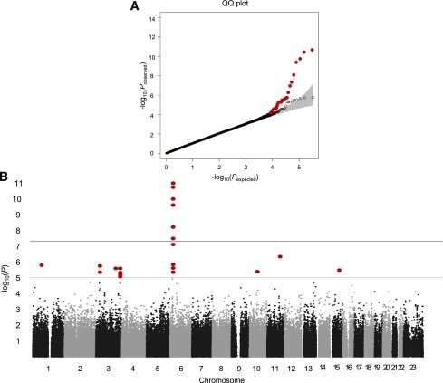
FIG. 1.
FIG. 1. Stage 1 genome scan results. A: QQ plot showing the distribution of the observed P values from the logistic regression analysis for the stage 1 genome scan against the expected distribution under the null hypothesis. The gray zone indicates the 95% CI. Colored circle, distribution of excess association signals driven by the known type 2 diabetes variants (listed in Table 4) and markers in LD (r2 > 0.8) with them. ○, distribution after excluding the known type 2 diabetes variants. B: Manhattan plot depicting the significance of all the association results of the stage 1 genome scan. SNP locations are plotted on the x-axis according to their chromosomal position. The negative log10 of P values derived from the logistic regression analysis under the additive model are plotted on the y-axis. (A high-quality color representation of this figure is available in the online issue.)
FIG. 2.
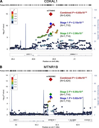
FIG. 2. Dense regional association plot near CDKAL1 (A) and MTNR1B (B). The hash marks above the panel represent the position of each SNP that was genotyped or imputed. The negative log10 of P values from logistic regression are shown in the panel. The blue diamond indicates the SNP with the most significant association in the stage 1 genome scan. The green and red diamonds represent the results of the SNP in stage 2 and joint stage 1 plus stage 2 analysis, respectively. Their corresponding P values are indicated on the right. Estimated recombination rates are plotted to reflect recombination hot spots. The SNPs in LD with the most significant SNP are color coded to represent their strength of LD. The locations of genes, exons and introns are shown in the lower panel (taken from the Human Genome hg18 build). (A high-quality color representation of this figure is available in the online issue.)
FIG. 3.
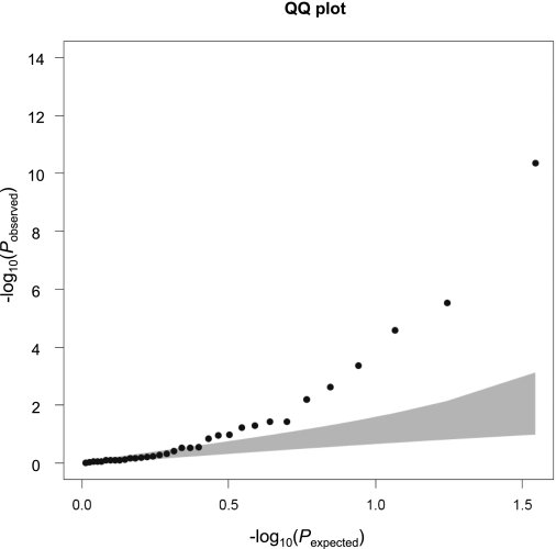
FIG. 3. QQ plot of the association between known type 2 diabetes variants and GDM. Comparison of the effect size of known type 2 diabetes variants in GDM and type 2 diabetes. The gray zone indicates the 95% CI. The known type 2 diabetes variants tested for association are listed in Table 4.
FIG. 4.
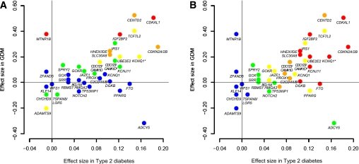
FIG. 4. Comparison of the effect size of known type 2 diabetes variants in GDM and type 2 diabetes. Effect size (β-coefficient from logistic regression analysis) of the known type 2 diabetes variants in GDM (y-axis) and type 2 diabetes (x-axis) are plotted with their corresponding P values (A: P values in GDM; B: P values in type 2 diabetes): red, P < 0.0001; orange, 0.0001 ≤ P < 0.01; yellow, 0.01 ≤ P < 0.10; green, 0.10 ≤ P < 0.50; blue, 0.50 ≤ P. The β-coefficient for GDM was derived from our stage 1 genome scan and that for type 2 diabetes was derived from the AGEN type 2 diabetes study (26). The two CDC123/CAMK1D variants are distinguished by CDC123/CAMK1D for rs10906115 and CDC123/CAMK1D* for rs12779790. The two KCNQ1 variants are distinguished by KCNQ1 for rs231362 and KCNQ* for rs2237892. (A high-quality color representation of this figure is available in the online issue.)

그림 출처: PubMed Central 공개 논문 (PMC3266417)

그 밖의 정보
출판 형태
Journal Article, Research Support, Non-U.S. Gov't
저널
Diabetes · eISSN 1939-327X
전자 출판
2012-01-10
MeSH
Adult · Aged · Cyclin-Dependent Kinase 5 / genetics · Diabetes Mellitus, Type 2 / genetics · Diabetes, Gestational / genetics · Female · Genome-Wide Association Study* · Humans · Insulin Resistance · Korea · Logistic Models · Middle Aged · Polymorphism, Single Nucleotide · Pregnancy · Receptor, Melatonin, MT1 / genetics · Risk Factors · tRNA Methyltransferases
Substances
Receptor, Melatonin, MT1 · tRNA Methyltransferases · Cyclin-Dependent Kinase 5 · CDKAL1 protein, human
참고문헌
48편