Department of Internal Medicine, Seoul National University College of Medicine, Seoul, Korea.
Abstract
The role and underlying mechanisms of rosiglitazone, a peroxisome proliferator-activated receptor-gamma (PPAR-γ) agonist, on myocardial infarction are poorly understood. We investigated the effects of this PPAR-γ agonist on the expression of tissue factor (TF), a primary molecule for thrombosis, and elucidated its underlying mechanisms. The PPAR-γ agonist inhibited TF expression in response to TNF-α in human umbilical vein endothelial cells, human monocytic leukemia cell line, and human umbilical arterial smooth muscle cells. The overexpression of TF was mediated by increased phosphorylation of mitogen-activated protein kinase (MAPK), which was blocked by the PPAR-γ agonist. The effective MAPK differed depending on each cell type. Luciferase and ChIP assays showed that transcription factor, activator protein-1 (AP-1), was a pivotal target of the PPAR-γ agonist to lower TF transcription. Intriguingly, two main drugs for drug-eluting stent, paclitaxel or rapamycin, significantly exaggerated thrombin-induced TF expression, which was also effectively blocked by the PPAR-γ agonist in all cell types. This PPAR-γ agonist did not impair TF pathway inhibitor (TFPI) in three cell types. In rat balloon injury model (Sprague-Dawley rats, n = 10/group) with continuous paclitaxel infusion, the PPAR-γ agonist attenuated TF expression by 70±5% (n = 4; P<0.0001) in injured vasculature. Taken together, rosiglitazone reduced TF expression in three critical cell types involved in vascular thrombus formation via MAPK and AP-1 inhibitions. Also, this PPAR-γ agonist reversed the paclitaxel-induced aggravation of TF expression, which suggests a possibility that the benefits might outweigh its risks in a group of patients with paclitaxel-eluting stent implanted.
Figures
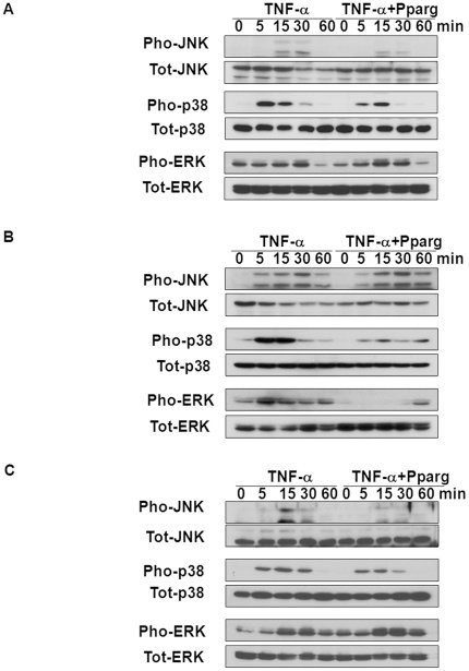
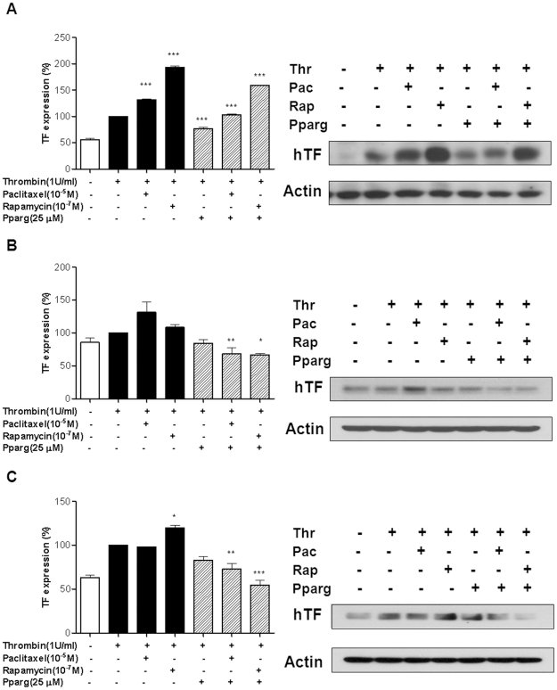
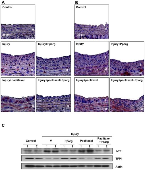
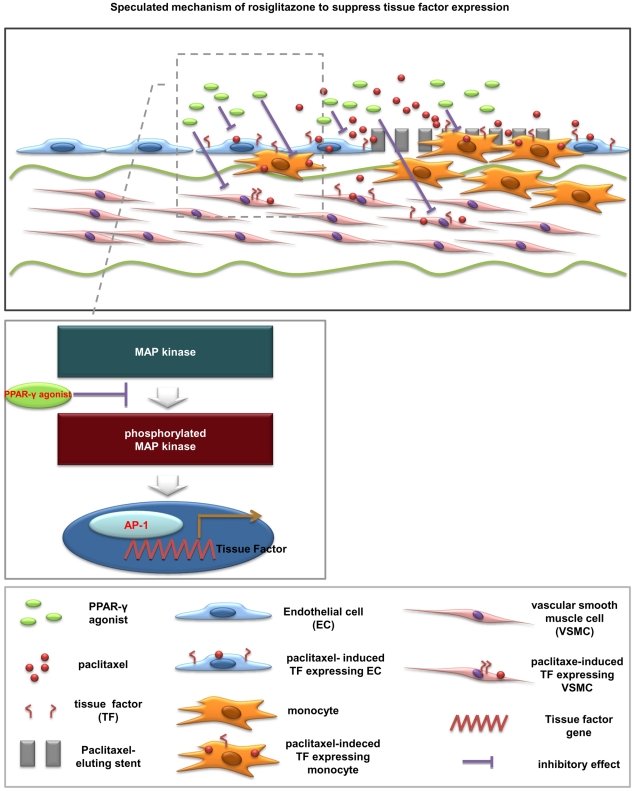
Figure 1 PPAR-γ agonist inhibited TF expression in ECs, monocytes and SMCs. (A) The PPAR-γ agonist suppressed TNF-α-induced TF expression in HUVECs in a concentration-dependent manner. (B–C) The PPAR-γ agonist also decreased TF expression in THP-1 (B) and SMCs (C). Average values of 3 different experiments. Data are presented as mean ± SEM. Values are given as percent of stimulation with TNF-α alone. *P = 0.002, **P<0.0001 vs TNF-α alone. Each number in western blot analysis indicates the concentration of the PPAR-γ agonist; all blots were normalized to actin expression. V = vehicle; Pparg = PPAR-γ agonist; hTF = human TF. See Figure S1 and S2 for the quantitative data and the effects of MAPK inhibitors on TF expression, respectively.Figure 2 Cell type-specific inhibition of MAPK phosphorylation. (A–C) The overexpression of TF was mediated by the increased phosphorylation (Pho) of MAPK, which was blocked by the PPAR-γ agonist. The main MAPK varied depending on each cell type; HUVECs were dependent on JNK and p38 (A); THP-1 cells were dependent on p38 and ERK (B); SMCs were dependent on JNK and p38 (C). Total (Tot) levels of MAPK remained unchanged.Figure 3 PPAR-γ agonist reduced TF mRNA expression in three cell types. (A–C) Real-time PCR demonstrated that TF mRNA levels increased after stimulation with TNF-α in HUVECs (A), THP-1 (B), and SMCs (C), which were suppressed by the PPAR-γ agonist. Data are presented as mean ± SEM. Values are given as percent of stimulation with TNF-α alone. *P<0.0001 vs TNF-α alone. All values are representative of 3 different experiments and are normalized to GAPDH. See also Figure S3.Figure 4 PPAR-γ agonist decreased TF promoter activity in three cell types. (A) The average fold induction of luciferase activity expressed by wild type or deletion mutant of TF promoter in response to the PPAR-γ agonist; three separate experiments were performed in triplicate (*P<0.05, **P<0.005, ***P<0.001). Data are presented as mean ± SEM. Luciferase assay demonstrated that AP-1 contributed to the inhibitory effect of the PPAR-γ agonist on TF expression in each cell type. (B) ChIP assay with AP-1 antibody verified that AP-1 was a critical transcription factor for TF-lowering effect of the PPAR-γ agonist in all cell types. See Figure S4 for the quantitative data.Figure 5 PPAR-γ agonist reversed the paclitaxel- or rapamycin-induced TF overexpression. (A–C) Paclitaxel and rapamycin aggravated thrombin-induced TF expression in HUVECs (A), THP-1 (B), and SMCs (C) (***P<0.0001 in HUVECs for paclitaxel or rapamycin vs thrombin alone; *P<0.05 in SMCs for rapamycin vs thrombin alone). The PPAR-γ agonist blunted the effect of paclitaxel and rapamycin on TF expression. Average values of 3 different experiments. Data are presented as mean ± SEM. Values are given as percent of stimulation with thrombin alone. *P<0.05, **P<0.01, ***P<0.0001 vs PPAR-γ agonist, PPAR-γ agonist plus paclitaxel, or PPAR-γ agonist plus rapamycin. Thr = thrombin; Pac = paclitaxel; Rap = rapamycin.Figure 6 The changes of TF and TFPI expressions after PPAR-γ agonist treatment in vivo. (A) Representative images of IHC staining for TF (hematoxylin-eosin stain, magnification x400). Specimens prepared from the injured and uninjured rat carotid arteries were examined. Balloon injury increased TF expression of injured arteries. TF expression was less prominent after the PPAR-γ agonist treatment than that from balloon injury alone. Paclitaxel infusion enhanced TF expression, which was reversed by the PPAR-γ agonist. (B) Representative images of IHC staining for TFPI (hematoxylin-eosin stain, magnification x400). The PPAR-γ agonist increased TFPI expression with or without paclitaxel. Brownish-red indicates TF (A) or TFPI protein (B). The upper region indicates luminal side. I = intima; M = media. (C) Western blotting of carotid artery lysates confirmed these effects of the PPAR-γ agonist on TF and TFPI expressions. See Figure S6 and S7 for the low-power view of the rat carotid arteries, and for the quantitative data, respectively.Figure 7 Speculated schematic illustration showing TF-lowering effect of PPAR-γ agonist in three cell types. In addition to acute coronary syndrome itself and endothelial denudation after stent implantation, paclitaxel induced thrombosis-prone condition by enhancing TF expression in ECs, monocytes and SMCs. Our study suggested that the PPAR-γ agonist suppressed TF expression in these cell types and this action is mediated by the inhibition of MAP kinase and AP-1 activity.
그림 출처: PubMed Central 공개 논문 (PMC3226685)
Conflict of interest statement
Competing Interests:The authors have declared that no competing interests exist.