Cancer-associated fibroblast-derived fibulin-5 promotes epithelial-mesenchymal transition in diffuse-type gastric cancer via cAMP response element-binding protein pathway, showing poor prognosis.
Division of Gastroenterology, Department of Internal Medicine and Liver Research Institute, Seoul National University College of Medicine, Seoul, Republic of Korea.
Department of Gastroenterology, Seoul National University Bundang Hospital, Seongnam, Republic of Korea.
Department of Pathology, Seoul National University College of Medicine, Seoul, Republic of Korea.
Liver Research Institute, Seoul National University College of Medicine, Seoul, Republic of Korea.
Center for Gastric Cancer, National Cancer Center, Goyang, Republic of Korea.
Department of Biomedical Sciences, BK21 FOUR Biomedical Science Project, and Medical Research Center, Seoul National University College of Medicine, Seoul, Republic of Korea.
Department of Surgery and Cancer Research Institute, Seoul National University College of Medicine, Seoul, Republic of Korea.
Department of Microbiology, Hanyang University College of Medicine, Seoul, Republic of Korea.
Division of Gastroenterology, Department of Internal Medicine and Liver Research Institute, Seoul National University College of Medicine, Seoul, Republic of Korea. crystal5@snu.ac.kr.
Liver Research Institute, Seoul National University College of Medicine, Seoul, Republic of Korea. crystal5@snu.ac.kr.
Abstract
Diffuse-type gastric cancer (DGC), characterized by poorly cohesive cells within fibrotic stroma, is associated with advanced disease and poor prognosis. Here, to identify distinct biomarkers for DGC compared with intestinal-type gastric cancer, we constructed a comprehensive large-scale signaling network using RNA-sequencing data from three genomic databases (The Cancer Genome Atlas, GSE62254 and GSE26253), developed a mathematical model and conducted simulation analyses. For validation, we used tissue microarray blocks of gastric cancers with immunohistochemical staining, single-cell RNA sequencing, primary cultures of cancer-associated fibroblasts (CAFs) and organoids, and a co-culture system involving CAFs and cancer cells. Signaling network analysis identified six differentially activated signaling components across the database, including BIRC5, TTK, NEK2, FHL1, NR2F1 and FBLN5. Among the differentially activated signaling components, high tumoral expression of fibulin-5 protein encoded by FBLN5 correlated with poor overall and disease-specific survival rates in patients with DGC, even after adjusting for the tumor, node, metastases (TNM) stage. Fibulin-5, derived from CAFs within DGC stroma, promoted organoid growth and epithelial-mesenchymal transition (EMT) in DGC cell lines via the cAMP response element-binding protein (CREB) pathway in a CAF co-culture system. FBLN5 knockdown in CAFs reduced the aggressive phenotype of co-cultured DGC cells, while CREB inhibitors reversed EMT. Furthermore, levels of secreted FBLN5 in patient blood samples correlated with its expression in primary tumors. In summary, fibulin-5 secreted by CAFs and interacted with DGC cells promotes EMT and is clinically associated with poor patient outcomes. These findings suggest fibulin-5 as a potential prognostic marker and therapeutic target in patients with DGC.
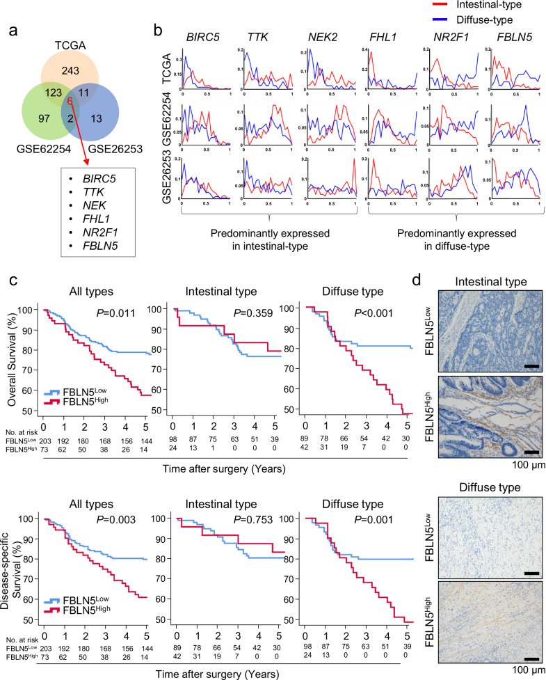
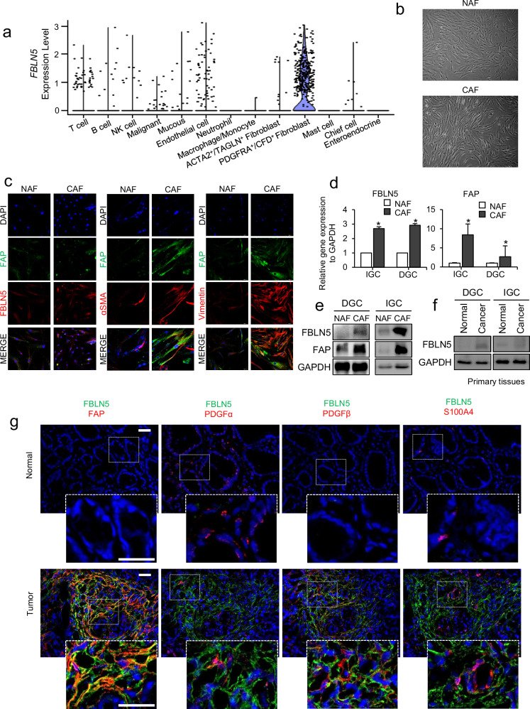
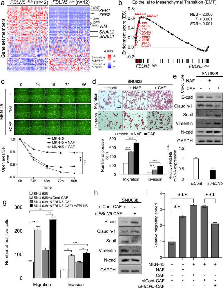
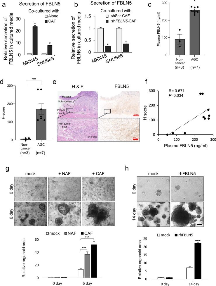
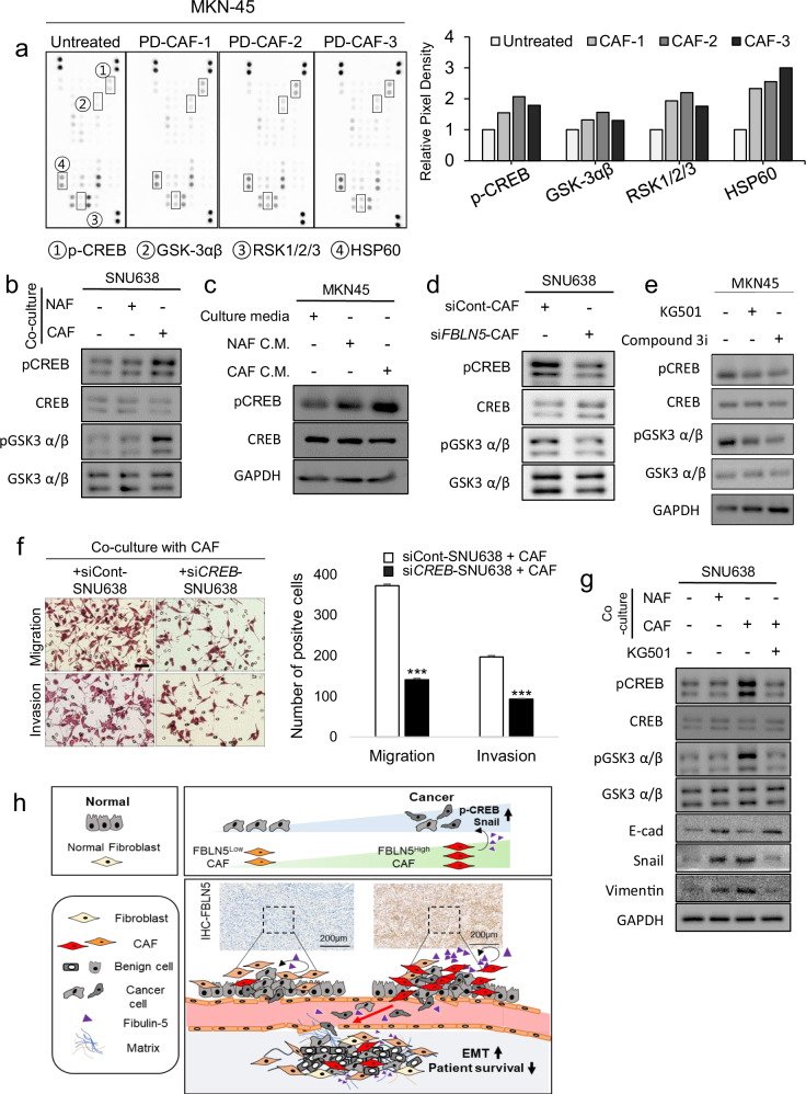
Fig. 1FBLN5 was one of the DASC distinguishing between IGC and DGC, which was highly activated in DGC and has worse prognostic outcome. a A large-scale network analysis utilizing data from three genomic databases (TCGA, GSE62254 and GSE26253) identified six DASCs common to all three databases. b The activity distribution of the six DASCs among IGC and DGC patients. The x axis represents the scale of activity while the y axis is the estimated density in distribution. The red line represents the density distribution of the signaling component in IGCs while the blue line represents that of DGCs. c Kaplan–Meier curves for OS (top) and DSS (bottom) stratified by low versus high expression of FBLN5 in all cancers and each Lauren subtype. d Representative IHC staining images of FBLNLow and FBLNHigh cancer in each Lauren subtype (intestinal type, top and diffuse type, bottom). Scale bar, 100 µm.Fig. 2 Fibulin-5 (FBLN5) is derived from CAFs in cancer stroma. a Single-cell RNA-sequencing data using four tissues from patients with DGC to investigate the origin of FBLN5. The dot plot shows the expression level of FBLN5 across 13 identified cell clusters, highlighting its predominant derivation from fibroblasts. b Microscopic images of isolated fibroblasts obtained by the outgrowth method from paired normal (top) and cancer (bottom) tissue of actual patients with DGC. c Verification of PD-NAFs and PD-CAFs is conducted through immunofluorescent staining for CAF markers and FBLN5. d The relative mRNA expression of FBLN5 and FAP is compared between PD-NAFs and PD-CAFs by RT–qPCR. *P < 0.05. e PD-NAFs and PD-CAFs, isolated from different Lauren DGC and IGC, are identified based on FAP and fibulin-5 expression levels through western blot analysis. f Fibulin-5 expression is compared in paired normal and primary tumor tissues from patients with DGC and IGC. g Immunofluorescent staining for CAF markers and fibulin-5 in paired normal and cancer tissues, accompanied by merged images showing the overlap of fibuilin-5 expression sites with CAF marker expression. DAPI (blue), FBLN5 (green) and CAF markers (FAP, PDGFα, PDGFβ and S100A4; red).Fig. 3 Fibulin-5 (FBLN5) promotes EMT of DGC. a A heat map generated from GSEA utilizing TCGA data illustrates the different gene distribution between FBLN5Low and FBLN5High patient cohorts. The mRNA expression levels, normalized via Z score transformation, indicate an enrichment of the EMT gene set in the FBLNHigh group relative to the FBLN5Low group. b Enrichment score plots (black curve) from GSEA for ranked genes within the EMT gene set reveal the NES, nominal P value and FDR. c Microscopic images of wound healing assays and corresponding line graphs depicting the relative open wound area over the cell area ratio at consecutive time points (hour) during live-cell imaging illustrate differences in wound healing between GFP-expressing MKN-45 stable cell line monoculture and co-cultures with PD-NAFs or PD-CAFs. ***P < 0.001. d A comparison of invasion and migration capabilities of the DGC cell line (SNU638) across cancer monoculture systems and co-culture systems with PD-NAFs and PD-CAFs represented through bar graphs. Scale bar, 50 µm. ***P < 0.001. e Western blot analysis of EMT markers in SNU638 cells under monoculture conditions and co-culture conditions with PD-NAFs or PD-CAFs. f Knockdown of FBLN5 using siFBLN5 in PD-CAFs co-culture systems compared with vehicle-treated PD-CAFs. *P < 0.02. g Invasion and migration assays of SNU638 cells in PD-CAF co-culture systems with or without siFBLN5 treatment reveal significant decrease of these capabilities on knockdown of FBLN5. Restoration of FBLN5 using rhFBLN5 in co-culture system with siFBLN5-CAF restores migration and invasion of cancer cells, which were decreased after the knockdown of FBLN5. **P < 0.01 and ***P < 0.001. h A reduction in EMT marker expression in DGC cells following FBLN5 knockdown in co-cultured CAFs compared with siControl (siCont) treatment in PD-CAFs is demonstrated. i Analysis of MKN-45 cell migration speed using the Operetta live-cell imaging system. Migration speed of GFP-expressing MKN-45 cells is compared under the conditions with the presence of patient-derived, NAFs, CAFs, siControl-treated CAFs and siFBLN5-treated CAFs. CAF-derived FBLN5 facilitates the migration of cancer cells, which was suppressed by siFBLN5 treatment. The bars represent average speed of MKN-45 (µm/s) and s.d. **P < 0.01 and ***P < 0.001.Fig. 4 Secreted fibulin-5 (FBLN5) from CAFs is detectable in the blood of patients, paralleling its expression in primary tissue. a A comparison of FBLN5 concentration in culture media of monocultures from DGC cell lines (MKN-45 and SNU668) and co-cultures of these cancer cells with induced CAFs (iCAFs). b The relative concentration change of FBLN5 in culture media from the co-culture system following to the knockdown of FBLN5 by transfection with shFBLN5 in iCAFs. c The concentrations of fibulin-5 (ng/ml), measured by ELISA in plasma samples, are compared between non-cancer subjects (n = 3) and patients with diffuse-type AGC (n = 7). *P < 0.05. d, The average H score, assessed from FBLN5 IHC staining, is compared between normal gastric tissues from non-cancer subjects (n = 3) and tumors from patients with AGC. **P < 0.01. e Representative paired H&E and IHC staining images for FBLN5 in AGC tissues display a gradient of expression from tumor to adjacent non-tumor tissues, indicative of local secretion and diffusion of FBLN. Scale bars, 2 mm and 500 µm (magnification ×10 and ×40). f Pearson’s correlation coefficient and scatter plot between the H score of gastric tissue and plasma FBLN5 concentration. g Microscopic examination of GCOs derived from an 83-year-old female patient with diffuse-type AGC shows different growth patterns among organoids cultured alone, with PD-NAFs and with PD-CAFs. Organoid areas were normalized to mean of day 0. The bar graph depicts the mean of individual organoid area per field for each group. Scale bar, 50 µm. ***P < 0.001. Scale bar, 50 µm. h Comparative microscopic images of GCOs treated with or without rhFBLN5 (10 ng/ml) illustrates an enhancement in organoid size with rhFBLN5 treatment, suggesting similar effect of CAF-derived FBLN5. Organoids area per field shown in bar graphs. ***P < 0.001. Scale bar, 50 µm.Fig. 5 CAF-derived fibulin-5 (FBLN5) increases cancer aggressiveness through CREB signaling. a A screening phosphokinase array to investigate the kinase pathway related to the downstream signal of fibulin-5 on DGC cells, co-cultured with patient-derived CAFs from three patients with diffuse-type AGC. The analysis reveals upregulation in the pixel density of four kinases (pCREB, GSK-3αβ, RSK 1/2/3 and HSP60), as depicted in the accompanying bar graph. b Representative western blot images display the differential expression of signaling components between the monoculture system and CAF co-culture system. The expression levels of CREB-GSK components (pCREB Ser133 and pGSK-3αβ Ser21/9) in the DGC cell line are elevated in co-culture systems with either NAFs or CAFs compared with monocultures. The lowest expression is observed in monoculture, followed by higher levels in co-culture with NAFs, and the highest expression in co-cultures with CAFs. c Western blot images shows the expression of CREB signal components across different culture conditions, including control conditioned media (CM), NAF-cultured CM and CAF-cultured CM. The expression of phosphorylated CREB (pCREB Ser133) is enhanced in conditions containing CAF-cultured CM, followed by NAF-cultured CM. d Western blot analysis following the knockdown of FBLN5 in CAFs reveals suppressed expression of CREB (pCREB Ser133) and GSK-3αβ (pGSK-3α Ser21/9) in the SNU 638 cells within the co-culture system. e The effects of CREB inhibitors (KG501 and Compound 3i (666-15)) on the MKN-45 cells are also assessed by western blot, showing decreased levels of phosphorylated CREB and phosphorylated GSK-3αβ upon treatment with these inhibitors. f Representative microscopic images from migration and invasion assays to evaluate the impact of CREB knockdown in DGC cells in a co-culture system with CAFs. The assays used SNU 638 cells and CAFs in co-culture systems with either a siControl or siCREB-treated DGC cell line. The number of migrating or invading cells are shown in the bar graphs. ***P < 0.001. Scale bar, 50 µm. g Western blot images of the expression of CREB-GSK signal components and EMT markers (E-cadherin, Snail and Vimentin) of SNU 638 cells co-cultured with either NAFs or CAFs. Treatment with CREB inhibitor KG501 reverses the effects induced by CAFs on SNU 638 cells. h An overview of the interplay between CAF-derived fibulin-5 and its influence on the aggressiveness of DGC through modulation of the CREB signaling pathway.
그림 출처: PubMed Central 공개 논문 (PMC12130531)
Conflict of interest statement
Competing interests: The authors declare no competing interests.
그 밖의 정보
출판 형태
Journal Article
저널
Experimental & molecular medicine · eISSN 2092-6413