Biomedical Research Institute, Seoul National University Hospital, Seoul, Republic of Korea.
Department of Molecular Medicine and Biopharmaceutical Sciences, Graduate School of Convergence Science and Technology, and College of Medicine or College of Pharmacy, Seoul National University, Seoul, Republic of Korea.
Program in Stem Cell Biology, Seoul National University College of Medicine, Seoul, Republic of Korea.
Department of Internal Medicine, Seoul National University Hospital, Seoul, Republic of Korea. hyunjaicho@snu.ac.kr.
Department of Internal Medicine, Seoul National University Hospital, Seoul, Republic of Korea.
Abstract
Lysophosphatidic acid receptor 4 (LPAR4) exhibits transient expression at the cardiac progenitor stage during pluripotent stem cell (PSC)-derived cardiac differentiation. Using RNA sequencing, promoter analyses, and a loss-of-function study in human PSCs, we discovered that SRY-box transcription factor 17 (SOX17) is an essential upstream factor of LPAR4 during cardiac differentiation. We conducted mouse embryo analyses to further verify our human PSC in vitro findings and confirmed the transient and sequential expression of SOX17 and LPAR4 during in vivo cardiac development. In an adult bone marrow transplantation model using LPAR4 promoter-driven GFP cells, we observed two LPAR4+ cell types in the heart following myocardial infarction (MI). Cardiac differentiation potential was shown in heart-resident LPAR4+ cells, which are SOX17+, but not bone marrow-derived infiltrated LPAR4+ cells. Furthermore, we tested various strategies to enhance cardiac repair through the regulation of downstream signals of LPAR4. During the early stages following MI, the downstream inhibition of LPAR4 by a p38 mitogen-activated protein kinase (p38 MAPK) blocker improved cardiac function and reduced fibrotic scarring compared to that observed following LPAR4 stimulation. These findings improve our understanding of heart development and suggest novel therapeutic strategies that enhance repair and regeneration after injury by modulating LPAR4 signaling.
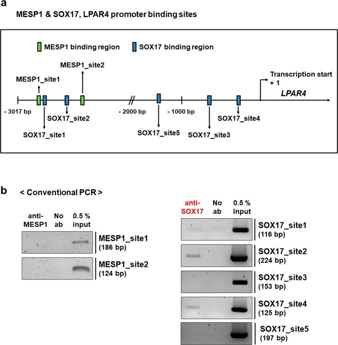
Fig. 1 Identification of the LPAR4 upstream transcription factor in hiPSC-derived cardiac differentiation. a Brightfield images during hiPSC-derived cardiac differentiation on Days 4 and 7 and in beating cardiomyocytes (CMCs). b RNA sequencing workflow for the three cell groups (n = 3). c Hierarchical cluster analyses of RNA sequencing during cardiac differentiation. d Clustering for the identification of LPAR4 upstream transcription factor candidates. e A heatmap showing candidate upstream transcription factors of LPAR4. f A schematic figure showing harvest days for real-time PCR and western blotting during hiPSC-derived cardiac differentiation. g Real-time PCR analysis of the expression of LPAR4 upstream transcription factor candidates during cardiac differentiation. Error bars represent the SEM, ***p < 0.001, unpaired t-test (n = 3 biological replicates). h Left, western blot analysis of LPAR4 expression of upstream transcription factor candidates during cardiac differentiation. Right, quantification of the obtained results (**p < 0.05, ***p < 0.001, means ± SEMs, three technical replicates). All experiments were conducted at least in triplicate.Fig. 2 Detection and binding sites of an LPAR4 upstream transcription factor. a Schematic showing the human MESP1 and SOX17 binding sites to the human LPAR4 promoter region targeted for chromatin immunoprecipitation (ChIP) assay and mutagenesis. Human LPAR4 promoter region of 3017 bp. b Binding of MESP1 or SOX17 to LPAR4 promoter regions using ChIP assays.Fig. 3 Mutation of the human LPAR4 promoter region to which human SOX17 binds and a gain or loss of function study for SOX17. a Schematic figure for SOX17 binding site mutations in the LPAR4 promoter region. A point mutation (C to A) in AACAAG(T), the consensus sequence that binds SOX17, was performed. Based on the ChIP assay results, mutation (mut.) sites 1 and 3 were used as negative controls, and site 5 was excluded because these sites do not bind SOX17, whereas mut. sites 2 and 4 are sites known to bind SOX17. A luciferase assay was performed with these point mutation constructs. b Activation of the human LPAR4 gene promoter in hiPSC-derived cells at cardiac differentiation Day 4 using the luciferase assay. ns; not significant, ***p < 0.001. All experiments were conducted at least in triplicate. c Real-time PCR analysis of mRNA expression levels of SOX17, LPAR4, and the early cardiac markers MESP1 and Nkx2.5 after cardiac differentiation using SOX17 knockdown (using SOX17 siRNA) hiPSCs. Statistical analyses were performed using one-way ANOVA (Newman‒Keuls). **p < 0.05, ***p < 0.001. d Real-time PCR analysis of mRNA expression levels of SOX17, LPAR4, Nkx2.5, and the mature cardiac marker αMHC after cardiac differentiation using SOX17-overexpressing hiPSCs (SOX17 OE). One-way ANOVA (Newman‒Keuls) was performed to assess significant differences (*p < 0.01, ***p < 0.001). All experiments were conducted at least in triplicate.Fig. 4 LPAR4 expression during mouse embryonic and postnatal development. a Schematic representation of LPAR4 lineage tracing mouse embryo harvesting (embryonic Day 9.5 (E9.5), E10.5, E12.5, and E16.5). At least three mouse embryos were harvested for each time point. b Immunostaining for GFP and α-SA expression during LPAR4 lineage tracing mouse embryonic development. Merged images for GFP (green) and α-SA (red) from whole mouse embryos (E9.5, E10.5, E12.5, and E16.5). Green, GFP; red, αSA; blue, DAPI (nuclei). Scale bar, 500 μm. c Immunofluorescence analysis of embryonic heart, liver, and BM from LPAR4 lineage tracing mice at E10.5 and E12.5. Green, GFP; red, Sca1; white, SOX17; blue, DAPI (nuclei). Scale bar, 10 μm. All experiments were conducted at least in triplicate. d FACS analysis of heart, liver, and BM cells from LPAR4 lineage tracing mouse neonates (postnatal day 1). FACS analysis was performed using GFP (FITC) for LPAR4 expression and SOX17 (APC) or Sca1 (PE) staining. e Immunofluorescence analysis of LPAR4 lineage tracing for mouse neonatal heart, liver, and bone marrow cells. Green, GFP; red, Sca1; white, SOX17, blue, DAPI (nuclei).Fig. 5 Characterization of LPAR4 in the myocardial infarcted heart after bone marrow transplantation. a Schematic figure of mouse bone marrow transplantation (BMT) and the myocardial infarction (MI) model. b FACS analysis of LPAR4 and GFP staining in non-MI hearts after BMT and in hearts on Days 4, 7, and 14 post-MI after BMT. c Top, line graph showing the percentage of LPAR4+GFP+ and LPAR4+GFP− cells in non-MI and post-MI hearts. Bottom, Venn diagram showing the ratio of LPAR4+GFP+ and LPAR4+GFP− cells in non-MI and post-MI hearts. d Representative flow cytometry plots of GFP-positive cells in the heart on Day 7 post-MI after BMT. Monocyte expression (CD11b+CD115+Ly6G−F4/80−), macrophage (CD11b+CD115−Ly6G−F4/80+), and neutrophil (CD11b+CD115−Ly6G+F4/80−) markers. e IF analyses were performed on hearts 4, 7, and 14 days after MI in BMT mice. BM-derived cells were stained with GFP (green), heart-resident LPAR4+ cells were stained with LPAR4 (red), and cardiac progenitor cells were stained with Nkx2.5 (white). Scale bar, 5 μm. f Quantification of the total percentage of Nkx2.5+ cells and LPAR4/Nkx2.5 double-positive cells in post-MI hearts. All experiments were conducted at least in triplicate.Fig. 6 Effect of treatment with a p38 MAPK blocker on cardiac differentiation and heart repair processes. a Echocardiography (sham heart group, n = 5; MI + PBS group, n = 4; MI + octadecenyl phosphate (ODP) [3 d] group, n = 8; MI + p38 MAPK blocker (SB203580) [3 d] group, n = 4; MI + ODP [3 d] + p38 MAPK blocker [3 d] group, n = 5). LVESD left ventricular end-systolic diameter, LVEDD left ventricular end-diastolic diameter, LVFS left ventricular functional shortening, LVEF left ventricular ejection fraction. b Hearts were fixed, sectioned, and MT stained. c Quantification of percent fibrotic area of MI hearts, analyzed by ImageJ. Statistical analyses were performed using one-way ANOVA (Newman‒Keuls). ns not significant, *p < 0.01, **p < 0.05, ***p < 0.001. Scale bar, 2 mm. All experiments were conducted at least in triplicate.
그림 출처: PubMed Central 공개 논문 (PMC10394006)
Conflict of interest statement
The authors declare no competing interests.
그 밖의 정보
출판 형태
Journal Article, Research Support, Non-U.S. Gov't
저널
Experimental & molecular medicine · eISSN 2092-6413