SCI 발표논문

SCI 발표논문

← SCI 발표논문 목록

Exp Mol Med. 2023;55(6):1174-1181.

Comparison of the effects of empagliflozin and sotagliflozin on a zebrafish model of diabetic heart failure with reduced ejection fraction.

, , , ,

제1저자 김인호 · 교신저자 석승혁, 이해영

소속 (9)
  1. Department of Internal Medicine, Seoul National University Hospital, Seoul, Korea.
  2. Department of Microbiology and Immunology, Seoul National University College of Medicine, Seoul, Korea.
  3. Department of Internal Medicine, Seoul National University College of Medicine, Seoul, Korea.
  4. Department of Internal Medicine, Seoul National University Bundang Hospital, Seongnam, Korea.
  5. Department of Microbiology and Immunology, Seoul National University College of Medicine, Seoul, Korea. lamseok@snu.ac.kr.
  6. Department of Biomedical Sciences, Seoul National University College of Medicine, Seoul, Korea. lamseok@snu.ac.kr.
  7. Cancer Research Institute, Seoul National University, Seoul, Korea. lamseok@snu.ac.kr.
  8. Department of Internal Medicine, Seoul National University Hospital, Seoul, Korea. hylee612@snu.ac.kr.
  9. Department of Internal Medicine, Seoul National University College of Medicine, Seoul, Korea. hylee612@snu.ac.kr.

Abstract

The sodium-glucose cotransporter 2 (SGLT2) inhibitor empagliflozin (EMPA) and dual SGLT1/2 inhibitor sotagliflozin (SOTA) are emerging as heart failure (HF) medications in addition to having glucose-lowering effects in diabetes mellitus (DM). However, the precise mechanism underlying this cardioprotective effect has not yet been elucidated. Here, we evaluated the effects of EMPA and SOTA in a zebrafish model of DM combined with HF with reduced ejection fraction (DM-HFrEF). To compare the effects of the two drugs, survival, locomotion, and myocardial contractile function were evaluated. The structural binding and modulating effects of the two medications on sodium-hydrogen exchanger 1 (NHE1) were evaluated in silico and in vitro. DM-HFrEF zebrafish showed impaired cardiac contractility and decreased locomotion and survival, all of which were improved by 0.2-5 μM EMPA or SOTA treatment. However, the 25 μM SOTA treatment group had worse survival rates and less locomotion preservation than the EMPA treatment group at the same concentration, and pericardial edema and an uninflated swim bladder were observed. SOTA, EMPA and cariporide (CARI) showed similar structural binding affinities to NHE1 in a molecular docking analysis and drug response affinity target stability assay. In addition, EMPA, SOTA, and CARI effectively reduced intracellular Na+ and Ca2+ changes through the inhibition of NHE1 activity. These findings suggest that both EMPA and SOTA exert cardioprotective effects in the DM-HFrEF zebrafish model by inhibiting NHE1 activity. In addition, despite the similar cardioprotective effects of the two drugs, SOTA may be less effective than EMPA at high concentrations.

© 2023. The Author(s).

Figures
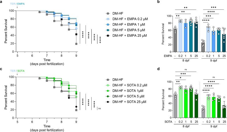
Fig. 1
Fig. 1 Comparison of survival rates in DM-HFrEF model zebrafish treated with empagliflozin or sotagliflozin. Survival analysis of DM-HFrEF zebrafish larvae after treatment with 0.2, 1, 5 and 25 μM empagliflozin or sotagliflozin. a, c Kaplan‒Meier survival curves (n = 120 larvae per group). b, d Ccross-sectional comparisons at the time points of 8 and 9 dpf and are presented as the mean ± standard deviation; each dot represents the value from one experiment (n = 3–6 experiments per group). The non-DM and DM groups were tested under the same conditions on the same day, and the graph is shown in Fig. 1 and Supplementary Fig. 2. *p < 0.05, **p < 0.01, ***p < 0.001, ****p < 0.0001 vs. the indicated group.
Fig. 2
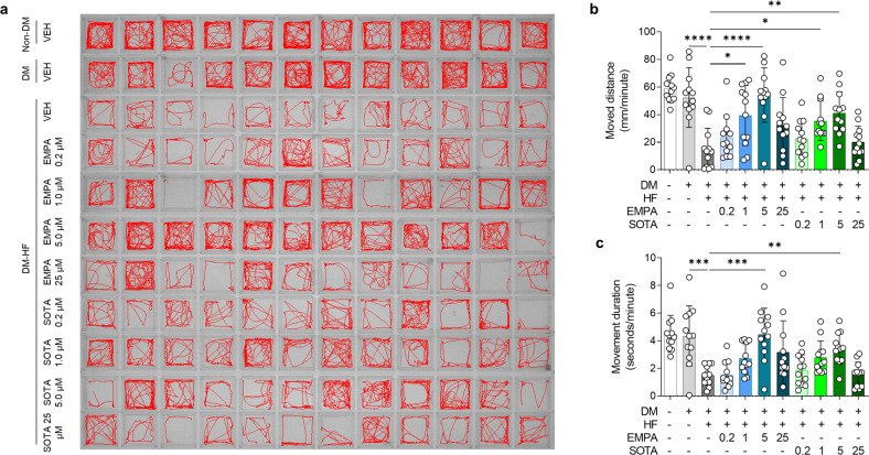
Fig. 2 Comparison of locomotion in DM-HFrEF zebrafish treated with empagliflozin or sotagliflozin. a Representative locomotion tracking images of zebrafish larvae for a 5-minute period. b, c Average movement distance and movement duration per 1 min (n = 12 larvae per group). Data are presented as the mean ± standard deviation, and each dot represents the value for one zebrafish larva. *p < 0.05, **p < 0.01, ***p < 0.001, ****p < 0.0001 vs. the indicated group.
Fig. 3
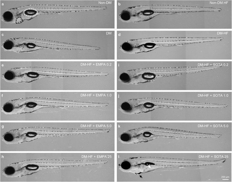
Fig. 3 Comparison of morphology in DM-HFrEF zebrafish treated with empagliflozin or sotagliflozin. Representative morphological images. a Non-DM, b non-DM with HFrEF, c DM, d DM-HFrEF. e–l DM-HFrEF zebrafish following treatment with e 0.2 μM, f 1 μM, g 5 μM and h 25 μM empagliflozin or i 0.2 μM, j 1 μM, k 5 μM, and l 25 μM sotagliflozin. a The pericardium is indicated by a black dashed line, the swim bladder by a white dashed line, l the pericardial edema by a black arrow, and uninflated swim bladder by a white arrow.
Fig. 4
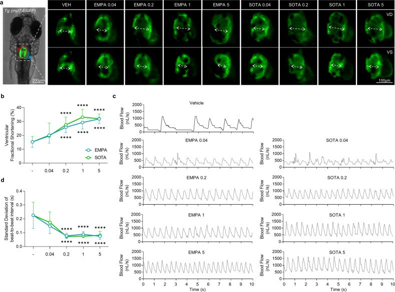
Fig. 4 Comparison of cardiac contraction in DM-HFrEF zebrafish treated with various concentrations of empagliflozin or sotagliflozin. a Representative fluorescence microscopy images of TG (myl7:EGFP) zebrafish hearts at end-diastole and end-systole of ventricle. b Ventricular fractional shortening calculated based on fluorescent images (n = 8–25 larvae per group). c Representative blood flow graphs. d Standard deviation of the beat-to-beat interval analyzed based on blood flow (n = 14–25 larvae per group). b, d Data are presented as the mean ± standard deviation. ****p < 0.0001 vs. control group.
Fig. 5
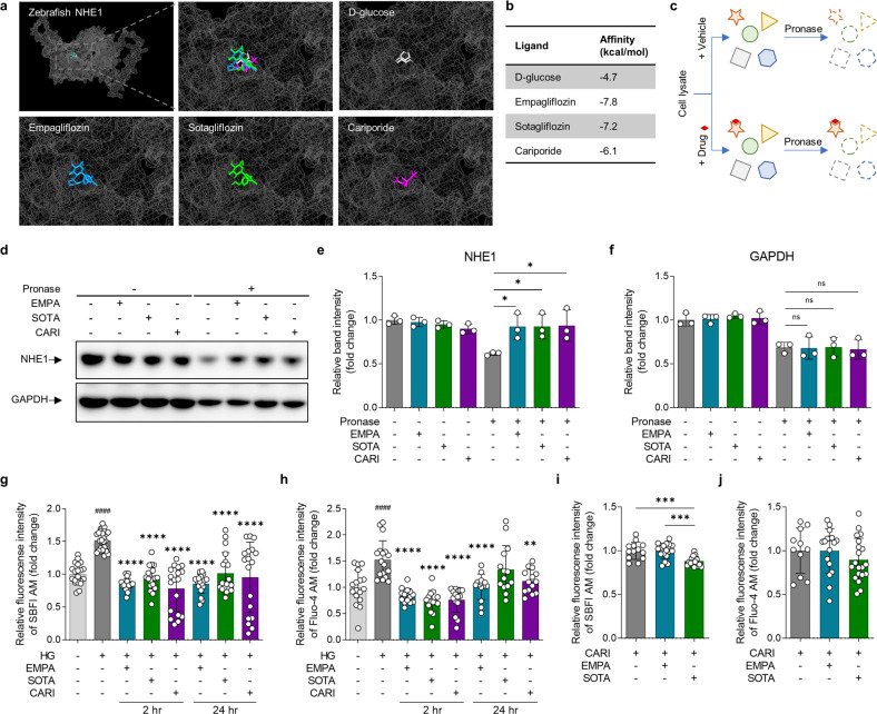
Fig. 5 Comparison of structural binding and functional inhibitory effects of empagliflozin and sotagliflozin on NHE1. a, b Molecular docking analysis of empagliflozin, sotagliflozin, cariporide and D-glucose binding to a structural model of zebrafish NHE1 in silico. Empagliflozin is shown in blue, sotagliflozin is shown in green, cariporide is in purple, and D-glucose is in gray. D-glucose was used as a negative control, and cariporide was used as a positive control. a Binding site and b affinities. c Schematic illustration of the drug affinity responsive target stability (DARTS) assay in vitro. d–f DARTS analysis by immunoblotting with NHE1 and GAPDH antibodies (n = 3 samples per group). g–j Measurement of intracellular Na+ and Ca2+ to analyze the functional inhibitory effects of empagliflozin and sotagliflozin on NHE1. g Changes in intracellular Na+ and h Ca2+ concentrations in cardiomyocyte under HG condition were evaluated after treatment with (n = 14–27 samples per group). i, j Changes in intracellular Na+ and Ca2+ concentrations in response to empagliflozin or sotagliflozin treatment in cells pretreated with cariporide (n = 11–20 samples per group). Data are presented as the mean ± standard deviation, and each dot represents the value of each sample. e, i *p < 0.05, ***p < 0.001 vs. the indicated group. g, h ####p < 0.0001 vs. the LG group, **p < 0.01, ****p < 0.0001 vs. the HG group.

그림 출처: PubMed Central 공개 논문 (PMC10318005)

Comment · 정정

Conflict of interest statement

The authors declare no competing interests.

그 밖의 정보
출판 형태
Journal Article, Research Support, Non-U.S. Gov't
저널
Experimental & molecular medicine · eISSN 2092-6413
전자 출판
2023-06-01
MeSH
Animals · Heart Failure* / metabolism · Zebrafish · Sodium-Glucose Transporter 2 Inhibitors* / pharmacology / therapeutic use · Stroke Volume · Molecular Docking Simulation · Diabetes Mellitus, Type 2* / drug therapy · Glucose · Benzhydryl Compounds · Glucosides · Glycosides · Guanidines · Sulfones
Substances
Sodium-Glucose Transporter 2 Inhibitors · Glucose · empagliflozin · Benzhydryl Compounds · Glucosides · (2S,3R,4R,5S,6R)-2-(4-chloro-3-(4-ethoxybenzyl)phenyl)-6-(methylthio)tetrahydro-2H-pyran-3,4,5-triol · Glycosides · cariporide · Guanidines · Sulfones
참고문헌
40편