Division of Pulmonary and Critical Care Medicine, Department of Internal Medicine, Seoul National University Hospital, Seoul, South Korea.
Department of Internal Medicine, Seoul National University College of Medicine, Seoul, South Korea.
Division of Pulmonary and Critical Care Medicine, Department of Internal Medicine, Seoul National University Hospital, Seoul, South Korea. cgyoo@snu.ac.kr.
Department of Internal Medicine, Seoul National University College of Medicine, Seoul, South Korea. cgyoo@snu.ac.kr.
Abstract
Inflammation, oxidative stress, and protease-antiprotease imbalance have been suggested to be a pathogenic triad in chronic obstructive pulmonary disease (COPD). However, it is not clear how proteases interact with components of inflammatory pathways. Therefore, this study aimed to evaluate the effect of neutrophil elastase (NE) on lipopolysaccharide (LPS)-induced interleukin 8 (IL-8) production and determine the molecular mechanism in human bronchial epithelial cells (HBECs). Immortalized bronchial epithelial cells and primary HBECs were used to investigate the impact of NE on LPS-induced IL-8 production. The molecular mechanism by which NE modulated LPS-induced IL-8 production was confirmed in elastase-treated C57BL/6 mice and primary HBECs obtained from COPD patients and healthy controls. The results showed that NE treatment synergistically augmented LPS-induced IL-8 production in both immortalized bronchial epithelial cells and primary HBECs. NE partially degraded peroxisome proliferator-activated receptor gamma (PPARγ), which is known to regulate IL-8 production in the nucleus. Treatment with a PPARγ agonist and overexpression of PPARγ reversed the NE-induced synergistic increase in LPS-induced IL-8 production. Moreover, PPARγ levels were lower in lung homogenates and lung epithelial cells from elastase-treated mice than in those from saline-treated mice. In accordance with the findings in mice, PPARγ levels were lower in primary HBECs from COPD patients than in those from healthy never-smokers or healthy smokers. In conclusion, a vicious cycle of mutual augmentation of protease activity and inflammation resulting from PPARγ degradation plays a role in the pathogenesis of COPD.
Figures
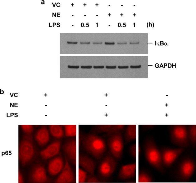
Fig. 1 NE synergistically augments the LPS-induced production of IL-8 in lung epithelial cells. BEAS-2B cells (a, b) and primary HBECs (c, d) were pretreated with VC or NE (1 U/mL) for 4 h and then stimulated with LPS (1 µg/mL) for 16 h in the presence or absence of VC or NE. Total RNA was isolated from BEAS-2B cells (a) and HBECs (c), and quantitative real-time PCR analysis of IL-8 and GAPDH was performed. The levels of IL-8 in the supernatants of BEAS-2B cells (b) and HBECs (d) were measured by ELISA. The data represent mean ± SD; **P < 0.05. Abbreviations: NE, neutrophil elastase; LPS, lipopolysaccharide; IL-8, interleukin 8; HBECs, human bronchial epithelial cells; VC, vehicle control; RNA, ribonucleic acid; PCR, polymerase chain reaction; GAPDH, glyceraldehyde 3-phosphate dehydrogenase; ELISA, enzyme-linked immunosorbent assays; SD, standard deviation.Fig. 2 NE does not affect LPS-induced degradation of IκBα or the subsequent nuclear translocation of the NF-κB subunit p65. a BEAS-2B cells were pretreated with VC or NE (1 U/mL) for 4 h and then stimulated with LPS (1 μg/mL) for the indicated times in the presence or absence of VC or NE. Total cellular extracts were subjected to western blot analysis for IκBα and GAPDH. b Cells were pretreated with either VC or NE and then incubated with LPS for 1 h in the presence or absence of VC or NE. The cells were fixed and permeabilized for 10 min. Immunofluorescent staining of p65 was performed using an anti-p65 primary antibody, followed by an Alexa Fluor 555 secondary antibody. Cells were analyzed using EVOS digital color fluorescence microscopy. Abbreviations: NE, neutrophil elastase; LPS, lipopolysaccharide; IκBα, nuclear factor of kappa light polypeptide gene enhancer in B-cells inhibitor alpha; NF-κB, nuclear factor kappa B; VC, vehicle control; GAPDH, glyceraldehyde 3-phosphate dehydrogenase.Fig. 3 NE downregulates PPARγ through proteolytic degradation. BEAS-2B cells were treated with VC or NE (1 U/mL) for the indicated times. a Total RNA was isolated, and quantitative real-time PCR analysis of PPARγ and GAPDH was performed. The data represent mean ± SD. b Total cellular extracts were subjected to western blot analysis for PPARγ and GAPDH protein quantification. c, d Cell lysates were incubated with VC or NE (0.00001, 0.0001, 0.001, 0.01, 0.25, and 1 U/mL) in the presence or absence of a serine protease inhibitor (PMSF, 1 mM) or an elastase inhibitor (Elaspol, 100 μg/mL) at 37 °C for 30 min. The reaction was terminated by adding an excess of sample buffer; subsequently, the samples were subjected to western blot analysis for PPARγ quantification. Abbreviations: NE, neutrophil elastase; PPARγ, peroxisome proliferator-activated receptor gamma; VC, vehicle control; RNA, ribonucleic acid; PCR, polymerase chain reaction; GAPDH, glyceraldehyde 3-phosphate dehydrogenase; SD, standard deviation; PMSF, phenylmethylsulfonyl fluoride.Fig. 4 Treatment with either a PPARγ agonist or overexpression of PPARγ suppresses the NE-induced increase in IL-8 production in LPS-treated cells. a BEAS-2B cells were pretreated with rosiglitazone (50 μM) for 1 h, followed by VC or NE (1 U/mL) for 4 h in the presence or absence of rosiglitazone. The cells were subsequently stimulated with LPS (1 μg/mL) for 24 h in the presence or absence of rosiglitazone. b, c BEAS-2B cells were transiently transfected with a pAdTrack-CMV-PPARγ expression vector or a control vector. Forty hours after transfection, the cells were pretreated with VC or NE (1 U/mL) for 4 h, followed by stimulation with LPS (1 μg/mL) for 16 h in the presence or absence of VC or NE. The concentrations of IL-8 in the supernatants were determined by ELISA (a, c). The data represent mean ± SD; **P < 0.05. Total cellular extracts were subjected to western blot analysis for PPARγ and GAPDH quantification (b). Abbreviations: PPARγ, peroxisome proliferator-activated receptor gamma; NE, neutrophil elastase; IL-8, interleukin 8; LPS, lipopolysaccharide; VC, vehicle control; ELISA, enzyme-linked immunosorbent assay; SD, standard deviation.Fig. 5 The expression levels of PPARγ are decreased in the lung epithelial cells of elastase-treated mice. a–c C57BL/6 mice were intratracheally administered either saline or elastase (0.5 U in 100 μL saline). The mice (n = 5 per group) were sacrificed 1 day after elastase treatment, and the lungs were isolated. Total lung protein lysates from the mice were subjected to western blot analysis for PPARγ and GAPDH quantification (a). The gel data were quantified using Scion image densitometry (b). The data represent mean ± SE; **P < 0.05. c PPARγ immunohistochemical analysis in lung tissues collected from mice. Arrows indicate epithelial cells stained with PPARγ. Abbreviations: PPARγ, peroxisome proliferator-activated receptor gamma; GAPDH, glyceraldehyde 3-phosphate dehydrogenase; SE, standard error.Fig. 6 PPARγ levels are decreased in the airway epithelial cells of COPD patients. a, b Large airway epithelial cells were obtained from never-smokers (n = 4), smokers (n = 4) and COPD patients (n = 4). c, d Small airway epithelial cells were acquired from never-smokers (n = 4), smokers (n = 8) and COPD patients (n = 11). Total cell lysates were subjected to western blot analysis for PPARγ and GAPDH quantification (a, c). The gel data were quantified using Scion image densitometry (b, d). The data represent mean ± SE; **P < 0.05. Abbreviations: PPARγ, peroxisome proliferator-activated receptor gamma; COPD, chronic obstructive pulmonary disease; GAPDH; glyceraldehyde 3-phosphate dehydrogenase; SE, standard error.
그림 출처: PubMed Central 공개 논문 (PMC8178386)
Conflict of interest statement
The authors declare no competing interests.
그 밖의 정보
출판 형태
Journal Article, Research Support, Non-U.S. Gov't
저널
Experimental & molecular medicine · eISSN 2092-6413